
Fluorescence Digital Image Gallery
Cells in Culture
Serious attempts at the culture of whole tissues and isolated cells were first undertaken in the early 1900s as a technique for investigating the behavior of animal cells in an isolated and highly controlled environment. The term tissue culture arose because most of the early cells were derived from primary tissue explants, a technique that dominated the field for over 50 years. As established cell lines emerged, the application of well-defined normal and transformed cells in biomedical investigations has become an important staple in the development of cellular and molecular biology. This fluorescence image gallery explores over 30 of the most common cell lines, labeled with a variety of fluorophores using both traditional staining methods as well as immunofluorescence techniques.
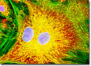
The bovine pulmonary artery endothelial (BPAE) cells presented in the digital image above were resident in an adherent culture stained for F-actin with Alexa Fluor 488 conjugated to phalloidin (green fluorescence), and for DNA with the bis-benzimidazole dye Hoechst 33258 (blue fluorescence). In addition, the culture was immunofluorescently labeled with Alexa Fluor 568 conjugated to goat secondary antibodies that target mouse anti-PDI (protein disulfide isomerase) primary antibodies (red fluorescence). The mitochondrial network was simultaneously visualized using MitoTracker Deep Red 633 (pseudocolored yellow). Images were recorded in grayscale with a Hamamatsu Orca-AG camera system coupled to an Olympus BX-51 microscope equipped with bandpass emission fluorescence filter optical blocks provided by Omega Filters. During the processing stage, individual image channels were pseudocolored with RGB values corresponding to each of the fluorophore emission spectral profiles.
African Green Monkey Kidney Cell Lines - The African green monkey has been a common subject of scientific inquiry for many years and cells from the tissues of this species, Cercopithecus aethiops, along with those of the rhesus monkey, have been used to produce polio vaccines since the 1950s. More recently, African green monkeys have been of significant interest because of the high levels of simian immunodeficiency virus type 1 (SIV-1) that frequently occur in their blood, which may be distantly related to the human immunodeficiency virus (HIV) that causes AIDS. In addition, several normal and transformed African green monkey kidney cell lines are excellent candidates for transfection investigations with recombinant plasmids.
Normal African Green Monkey Kidney Fibroblast Cells (CV-1) - The CV-1 cell line was initiated in March of 1964 by F. C. Jensen and his colleagues with a tissue section excised from the kidney of a normal adult male African green monkey (Cercopithecus aethiops). The popular fibroblast line was originally utilized in research focusing on the transformation of the cancer-causing Rous sarcoma virus (RSV), but now is a very useful host for acquired immunodeficiency disease (AIDS) research, as well as transfection experiments with simian virus 40 and recombinant plasmid vectors.
Normal African Green Monkey Kidney Epithelial Cells (Vero) - The Vero epithelial cell line was established in 1962 by Y. Yasumura and Y. Kawakita at the Chiba University in Chiba, Japan. The tissue from which the line was derived was obtained from the kidney of a healthy adult African green monkey. Although widely used in transfections and vaccine production, Vero cells are also often utilized in the detection of verotoxins, a group of interrelated toxins produced by some strains of Escherichia coli that are a key cause of hemorrhagic colitic and hemolytic uremic syndrome in humans.
Transformed (Simian Virus 40) African Green Monkey Kidney Fibroblast Cells (COS-1) - The transformed COS-1 cell line was derived from the CV-1 fibroblast line, which was initiated in March, 1964 by F. C. Jensen and colleagues from the normal kidney of an adult African green monkey. Developed by Yakov Gluzman, the COS-1 cell line differs from CV-1 due to transformation of the earlier line with an origin defective mutant of simian virus 40 (SV40) that codes for wild type T-antigen. Two other transformed lines, COS-3 and COS-7, were also established by Gluzman, but COS-1 cells are unique in the fact that they contain a single integrated copy of the complete early region of SV40 DNA. All three of the lines, however, fully support the lytic growth of SV40 (tsA209 strain) at 40 degrees Celsius and the replication of SV40 mutants with deletions in the early region.
Transformed (Simian Virus 40) African Green Monkey Kidney Fibroblast Cells (COS-7) - The COS-7 cell line was derived by Yakov Gluzman in the early 1980s from the previously established CV-1 African green monkey kidney line by transformation of the normal cells with an origin defective mutant of simian virus 40 (SV40) that codes for the wild-type virus T-antigen. The fibroblast line grows adherently to glass and plastic in culture and is generally utilized as a transfection host for virus genomes and recombinant plasmids.
African Water Mongoose Skin Fibroblast Cells (APM) - The APM cell line was established at The Naval Biosciences Laboratory (NBL) in Oakland, California from the skin of an African water mongoose (Atilax paludinosus). An elusive animal, the African water mongoose exhibits solitary, nocturnal habits and is a good swimmer, although it frequently lingers in shallow waters where it catches shellfish, crabs, frogs, and similar aquatic and semi-aquatic animals for food. APM mongoose cells exhibit fibroblast morphology and, similar to other fibroblast lines, are among the easiest cells to grow in culture. Cell biologists hypothesize that the ability of fibroblasts to grow so readily outside of the body is associated with their central role in the healing of wounds, which necessitates their proliferation when confronted with injury or other less than optimal conditions. Fibroblasts are also generally considered to exhibit relatively solitary lifestyles, which some have suggested may also be a factor in their favorable growth in culture.
Baby Hamster Kidney Fibroblast Cells (BHK-21) - The BHK-21 fibroblast cell line was established in March of 1961 by I. A. Macpherson and M. G. P. Stoker. The widely used line is a subclone (clone 13) of a parental line derived from the kidneys of five unsexed, 1-day-old hamsters. These hamsters were of the species Mesocricetus auratus and are commonly known as Syrian golden hamsters. Subsequent to 84 days of continuous cultivation, interrupted only for an 8-day preservation by freezing, clone 13 was initiated by single-cell isolation. BHK-21 cells are susceptible to human adenovirus D, reovirus 3, and vesicular stomatitis virus (Indiana strain), but are resistant to poliovirus 2. In addition, the cells are negative for reverse transcriptase, indicating the lack of integral retrovirus genomes. The BHK-21 line has been utilized as a host for transformation with expression vectors containing selectable and amplifiable marker DNAs and is also useful for transfections.
Bovine Pulmonary Artery Endothelial Cells (BPAE) - The BPAE cell line was initiated in January 1979 by P. Del Vecchio from the main stem of a pulmonary artery belonging to a young cow (Bos taurus). Pulmonary arteries, which extend from the heart to the lungs, are the only arteries in the mammalian body that carry dark, unoxygenated blood. The BPAE line of endothelial cells is positive for bovine diarrhea virus, one of the most important known bovine viral pathogens, which causes a broad array of clinical syndromes that result in significant losses in the beef industry each year. BPAE cells are also positive for angiotensin converting enzyme (ACE), an enzyme that is intricately involved in the maintenance of blood pressure and volume. Due to this fact, BPAE cells are often utilized in hypertension research as well as studies of atherosclerosis and coronary heart disease.
Embryonic Rat Thoracic Aorta Medial Layer Myoblast Cells (A-10) - The clonal cell line A-10 was derived by B. Kimes and B. Brandt from the thoracic aorta of an embryonic rat (Rattus norvegicus) from the established strain DB1X. The thoracic aorta is a branch of the descending aorta, which transports blood from the heart to the other organs and parts of the body. This arbitrary anatomic entity is generally considered to extend from the arch of the aorta to the diaphragm. The cells grow adherently and exhibit myoblast morphology, possessing many of the properties characteristic of smooth muscle cells. Cellular products include myokinase, creatine phosphokinase, and myosin. A-10 cells produce spontaneous action potentials at the stationary phase of the growth cycle and exhibit an increase in activity of the enzymes myokinase and creatine phosphokinase.
Embryonic Swiss Mouse Fibroblast Cells (3T3) - Established by George Todaro and Howard Green in 1962 from disaggregated Swiss mouse (Mus musculus) embryo tissue, the 3T3 cell line is a standard fibroblast cell line used in a wide spectrum of research and industrial biomedical applications. Variants of the initial cell line have been tested and found negative for ectromelia virus (mousepox), but most are susceptible to polyoma and simian virus 40 (SV40). In addition, 3T3 cells are negative for the viral enzyme reverse transcriptase, indicating the lack of integral retrovirus genomes. Within the cytoplasm, lysophosphatidylcholine (lyso-PC) induces AP-1 activity and c-jun N-terminal kinase activity (JNK1) by a protein kinase C-independent pathway. Contact inhibited, a confluent monolayer of 3T3 cells yields approximately 40,000 cells per square centimeter.
Female Rat Kangaroo Kidney Epithelial Cells (PtK1) - The PtK1 cell line was derived in 1962 from the kidney of a normal adult female Potorous tridactylus, a species of rat kangaroo prevalent in Australia. The epithelial line is believed to be the first permanent cell line of marsupial origin to be established. PtK1 cells are positive for keratin by immunoperoxidase and fluorescence staining and are susceptible to vesicular stomatitis (Indiana strain). PtK1 epithelial cells are resistant, however, to poliovirus 2 and are also negative for reverse transcriptase, indicating the lack of integral retrovirus genomes. The cell line has been primarily utilized in mitotic research because of the low number, large size, and distinct morphology of the chromosomes of the species from which it was established.
Horse Dermal Fibroblast Cells (NBL-6) - The NBL-6 cell line, also known as E. Derm, was derived from the dermis of a 4-year-old female horse (Equus caballus) of the quarterhorse strain. The cells, which appear to senesce after approximately 40 passages, are susceptible to herpes simplex, reovirus 3, vesicular stomatitis (Ogden strain), and vaccinia, but resist adenovirus 5, coxsackievirus A9 and B5, and poliovirus 2. NBL-6 cells, which are negative for reverse transcriptase and exhibit typical fibroblast morphology, have been utilized for a variety of studies, as well as for virus propagation for equine vaccines. The cell line has been particularly important in research focusing upon equine viral arteritis (EVA), a contagious viral disease that affects horse populations worldwide and appears to be increasing in incidence.
Human Bone Osteosarcoma Cells (U-2 OS) - The U-2 OS cell line, originally known as the 2T line, was derived from the bone tissue of a fifteen-year-old human female suffering from osteosarcoma. Established by J. Ponten and E. Saksela in 1964, the original cells were taken from a moderately differentiated sarcoma of the tibia. U-2 OS cells exhibit typical epithelial morphology and viruses were not detected in the line during co-cultivation with WI-38 cells or in CF tests against simian virus 40 (SV40), respiratory syncytial virus (RSV), or adenoviruses. Mycoplasma contamination in major stocks of the U-2 OS line was detected and subsequently eliminated in 1972. Cells are positive for insulin-like growth factor I (IGF-I) and insulin-like growth factor II (IGF II) receptors and express a number of antigens, including blood type A, Rh+, HLA A2, Aw30, B12, Bw35, and B40(+/-).
Human Brain Glioma Cells (U-118 MG) - The U-118 MG cell line is one of several cell lines derived from malignant gliomas by J. Ponten and associates in the late 1960s. The source for the cells of this particular line was a 50-year-old Caucasian male. The morphology of U-118 MG line is mixed and both glioblastoma and astrocytoma cells are present. U-118 MG cells are very similar to the glioblastoma U-138 MG cell line, though the line is supposed to have been derived from a different source. Studies have found that both of these lines have identical VNTR (variable number of tandem repeats) patterns and similar STR (short tandem repeat) patterns in DNA analysis experiments. They also have in common at least six derivative marker chromosomes. Mycoplasma contamination was detected and eliminated from the U-118 MG cell line in 1987 through treatment of cultures with BM-cycline over a six-week period. The cells have been demonstrated to be tumorigenic in nude mice subcutaneously inoculated.
Human Cervical Adenocarcinoma Cells (HeLa) - The HeLa line is one of the best-known cell lines in the world. Derived in 1951 from an adenocarcinoma of the cervix found in a 31-year-old woman (Henrietta Lacks), HeLa cells were the first human cells to survive indefinitely in the laboratory. The cells exhibit epithelial morphology and grow adherently, reproducing an entire generation about every 24 hours. Cellular products of the HeLa line include keratin and lysophosphatidylcholine (lyso-PC), which induces AP-1 activity and c-jun N-terminal kinase activity (JNK1) by a protein kinase C-independent pathway. HeLa cells have been reported to contain human papilloma virus 18 (HPV-18) sequences. P53 expression in the cells has been described as low, though levels of pRB (retinoblastoma suppressor) are apparently normal.
Human Cortical Neuronal Cells (HCN-1A) - The source for the HCN-1A cell line was cortical tissue removed from a patient undergoing hemispherectomy for intractable seizures. The patient was an 18-month-old female suffering from unilateral megalencephaly. Also known as hemimegalencephaly, this condition is characterized by the overgrowth of all or part of one of the cerebral hemispheres. HCN-1A cells stain positively for a number of neuronal markers including neuron specific enolase. They are also positive for tubulin, vimentin, somatostatin, glutamate, gamma aminobutyric acid, cholecystokinin-8, and vasoactive intestinal peptide. The cells are negative, however, for glial fibrillary acidic protein and myelin basis protein. HCN-1A cells can be induced to differentiate when cultured with a mixture of nerve growth factor, dibutyryl cyclic adenosine monophosphate, and 1-isobutyl-3-methylxanthine. Differentiation is accompanied by mature morphology and a deceleration of growth.
Human Fetal Lung Fibroblast Cells (MRC-5) - The MRC-5 cell line is commonly utilized in vaccine development, as a transfection host in virology research, and for in vitro cytotoxicity testing. Initiated in September 1966 by J. P. Jacobs, the cell line was derived from the human lung tissue of a 14-week-old male fetus aborted from a 27-year-old woman. MRC-5 cells, which grow adherently in culture and exhibit fibroblast morphology, may double in population size 42 to 46 times before the onset of senescence. They are susceptible to poliovirus 1, herpes simplex, and vesicular stomatitis (Indiana strain). The line is, however, negative for reverse transcriptase, indicating the lack of integral retrovirus genomes.
Human Lung Carcinoma Cells (A-549) - The A-549 cell line was originally cultivated in 1972 by D. J. Giard, along with several collaborators, from the human lung carcinoma of a 58-year-old Caucasian male. The line is commonly used to investigate a wide range of respiratory ailments, such as viral infections capable of inducing asthma, tissue damage linked to asbestos exposure, and smoking-related emphysema. Adherent and epithelial, A-549 cells are positive for keratin by immunoperoxidase staining, but are negative for reverse transcriptase, indicating the lack of integral retrovirus genomes. Studies by a team led by M. Lieber have revealed that A-549 cells are able to synthesize lecithin with a high percentage of desaturated fatty acids utilizing the cytidine diphosphocholine pathway.
Human Skin Epidermoid Carcinoma Epithelial Cells (A-431) - The A-431 cell line, which was derived from an epidermoid carcinoma excised from the skin tissue of an 85-year-old female, is one of a succession of cell lines established from solid tumors by a research team led by D. J. Giard. The epithelial line is tumorigenic, forming rapidly growing subcutaneous tumors in immunosuppressed mice and colonies in soft agar. Also known as cutaneous squamous cell carcinoma, epidermoid carcinoma of the skin is a malignant tumor of epidermal keratinocytes, a condition that has increased in frequency considerably over the last century, perhaps due in part to the ongoing depletion of the protective ozone layer.
Indian Muntjac Deer Skin Fibroblast Cells - A fibroblast cell line established from a skin biopsy of an adult male, the Indian Muntjac deer epidermis line is commonly used in laboratories around the world, especially for chromosome studies. Members of the family Cervidae, Muntjacs are barking deer that emit their characteristic sound when they feel threatened or alarmed. The normal (non-transformed) Indian Muntjac cell line is susceptible to the herpes simplex virus, vaccinia virus, and vesicular stomatitis virus (Indiana strain), but is resistant to poliovirus 1. Recent tests have demonstrated that the cells produce both detectable bovine viral diarrhea virus (BVDV) antigens and infectious BVDV virions. Muntjac cells are negative for reverse transcriptase, indicating the lack of integral retrovirus genomes.
Madin-Darby Canine Kidney Epithelial Cells (MDCK) - Derived by S. H. Madin and N. B. Darby from the kidney tissue of an adult female cocker spaniel, the MDCK cell line originated in September 1958. Since that time, the cells have been widely utilized to investigate the processing of beta-amyloid precursor protein, as well as the sorting of its proteolytic products. The morphology of the MDCK cell line is epithelial, and the cells are positive for keratin by immunoperoxidase staining. Viruses that MDCK cells are susceptible to include vesicular stomatitis (Indiana strain), vaccinia, coxsackievirus B5, reovirus 2 and 3, adenovirus 4 and 5, vesicular exanthema of swine, and infectious canine hepatitis. The cells exhibit resistance to coxsackievirus B3 and B4 as well as poliovirus 2, and are negative for reverse transcriptase.
Madin-Darby Ovine Kidney Epithelial Cells (MDOK) - The Madin-Darby ovine kidney (MDOK) cell line was derived from the renal tissue of a male sheep. The line exhibits typical epithelial morphology and is susceptible to several viruses including vesicular stomatitis (Indiana and New Jersey strains), infectious bovine rhinotracheitis, and sheep bluetongue virus. Epithelial cells generally exist in the body in sheets covering the organs and other internal and external surfaces that may come into contact with foreign materials. Usually the cells contain a relatively large amount of cytoplasm and a significant quantity of granules. The function of the cells is various, some acting in an absorptive or protective role, while others primarily act as secretory cells.
Male Rat Kangaroo Kidney Epithelial Cells (PtK2) - The widely used PtK2 cell line was established from the kidney tissue of an adult male rat kangaroo (Potorous tridactylus), a long-nosed, marsupial that is fairly common in Australia. The epithelial line is positive for the tough, insoluble intermediate filament protein keratin by immunoperoxidase staining, but is negative for reverse transcriptase, indicating the lack of integral retrovirus genomes. PtK2 cells are susceptible to coxsackievirus A9, herpes simplex, vaccinia, and vesicular stomatitis (Ogden strain). The cells are resistant, however, to adenovirus 5, coxsackievirus B5, and poliovirus 2. The PtK2 epithelial cell line is utilized for a variety of applications, but primarily for research in the field of mitosis.
Mink Uterus Endometrium Epithelial Cells (GMMe) - The GMMe cell line was established by the stable transfection of endometrial tissue of an adult female mink (Mustela vison) using a plasmid vector encoding the simian virus 40 (SV40) large T-antigen driven by the human beta-actin promoter. The cells were cotransfected with a second plasmid vector to confer neomycin resistance and were selected in medium containing G418. The cuboidal GMMe cells, which exhibit epithelial characteristics, are strongly positive for cytokeratin and weakly positive for vimentin. The line is also positive for alkaline phosphatase, but negative for desmin. GMME cells have been used in co-culture with mink embryos in obligate diapause to enhance the length and frequency of embryo survival in vitro, although the mink stromal line, GMMs, was utilized in this same manner with a greater rate of success.
Mouse Embryo Teratocarcinoma Epithelial Cells (P19) - The P19 cell line was derived from an embryonal carcinoma induced in a C3H/He mouse strain of the species Mus musculus. Similar to other embryonal carcinoma cells, cells of the P19 line are capable of differentiating into an array of cell types and are, therefore, described as pluripotent. Though they can be maintained and propagated in tissue culture in an undifferentiated state, when exposed to 500 nM of retinoic acid, P19 cells can be induced to differentiate into neural and glial-like cells. In the presence of 0.5 to 1.0 percent dimethylsulfoxide (DMSO), however, the cells differentiate to form cardiac and skeletal muscle-like elements, but do not form neural or glial-like cells. When introduced simultaneously to both DMSO and retinoic acid, P19 cells differentiate as if they were exposed only to retinoic acid.
Mouse Hemangioendothelioma Endothelial Cells (EOMA) - The EOMA cell line was derived in 1980 from a mixed hemangioendothelioma present in an adult mouse (Mus musculus). Hemangioendothelioma is the term utilized to describe a varied group of vascular tumors that usually appear as red or blue nodules and tend to behave biologically in a manner that can be classified as falling between a benign hemangioma and malignant angiosarcoma. EOMA cells are tumorigenic in syngeneic mice and exhibit characteristic endothelial cell properties. The cells synthesize a number of cellular products including angiotensin-converting enzyme (ACE), thrombospondin, Cathepsin L, endostatin, and interleukin-6. Surface receptors for acetylated low-density lipoprotein are expressed by EOMA cells, as is the vascular addressin, a tissue-specific endothelial cell adhesion molecule identified by antibody MECA-99.
Normal Rat Kidney Epithelial Cells (NRK) - The NRK cell line was derived from the kidney of a rat of the species Rattus norvegicus. The cells grow adherently and exhibit epithelial morphology. Epithelial cells are cells that exist in the body in sheets covering the organs and other internal and external surfaces that may come into contact with foreign materials. Typically epithelial cells contain a relatively large amount of cytoplasm and a significant quantity of granules. The function of the cells is various, some acting in an absorptive or protective role, while others primarily act as secretory cells. The epithelial cells of the kidneys chiefly fall into this latter category, playing an important role in the storage and subsequent secretion of various excretory materials.
Pig Kidney Epithelial Cells (LLC-PK1) - The LLC-PK1 cell line was derived from the kidney of a normal, healthy male pig (Sus scrofa) that was between 3 and 4 weeks of age. The pig was a member of the Hampshire breed, which was initially imported to America from Hampshire County, England in the early 1800s. Commonly utilized in laboratories around the world, the LLC-PK1 line exhibits typical epithelial morphology. Cellular products of the LLC-PK1 porcine kidney line include plasminogen activator, a substance that stimulates fibrinolysis. In recent years, plasminogen activator has been included in drugs used in thrombosis therapies since it facilitates the dissolution of small blood clots. The cells also produce large amounts of cytokeratin. The LLC-PK1 line is often used as a model for epithelial tissue, as well as in a wide spectrum of pharmacologic and metabolic research investigations.
Rat Jejunum Myenteric Plexus Enteroglial Cells (EGC/PK060399egfr) - Enteroglial cells (known by the acronym: EGC) are believed to be an important part of the enteric nervous system, but much about the function of these cells is still unknown. To help facilitate future studies of the cells, A. Ruhl and coworkers developed a new method in 2001 for isolating and purifying enteroglial cells from the myenteric plexus, a network of nerve fibers located in the intestinal wall. The Ruhl method involved enzymatic dissociation of myenteric plexus samples, the purification of enteric glial cells via complement-mediated cytolysis of contaminating cells, and transformation by retroviral gene transfer. The group then characterized the resulting clones both immunohistochemically and by dot-blot analysis. As a result of their efforts, a number of transformed EGC lines that retain their glial and functional characteristics have been established.
Rat Kidney Mesangial Cells (RMC) - The RMC cell line was derived from the kidney tissue of a 3-month-old male rat (Rattus norvegicus) belonging to the Sprague-Dawley strain. The mesangial cells were immortalized at passage eight with the plasmid pSV3-Neo, which codes simian virus 40 (SV40) large T-antigen, and are maintained in the presence of the antibiotic, G-418. The rat kidney cells express normal genes of the wild type mesangial cells and grow adherently to glass and polymer surfaces in monolayer culture. RMC cells are positive for desmin and vimentin, but are negative for cytokeratin 8. Mesangial cells are specialized cells usually associated with glomeruli that are crucial for kidney function. Thus, the RMC line and other lines of mesangial cells are widely utilized in kidney research.
Tahr Ovary Epithelial Cells (HJ1.Ov) - The HJ1.Ov cell line was derived from the ovary tissue of a normal and healthy female Himalayan tahr (Hemitragus jemlahicus), which is a relative of the wild goat specially adapted to life in the rugged, mountainous environment of the Himalayas. Developed at The Naval Biosciences Laboratory (NBL) in Oakland, California, continuous cultures of HJ1.Ov cells exhibit typical epithelial morphology and grow adherently to glass and polymer surfaces in monolayer culture. Epithelial cells comprise the avascular tissues that line both the interior and exterior surfaces of the body and its organs. Also, the secretory portions of glands and their ducts are formed from invaginated epithelial cells. Though there are many different types of epithelial cells in the body that may be arranged in a number of ways, the cells are always contiguous with one another so that they create an uninterrupted barrier.
Transformed Chicken Embryo Fibroblast Cells (UMNSAH/DF-1) - UMNSAH/DF-1 is a spontaneously immortalized chicken (Gallus gallus) cell line derived from 10-day-old East Lansing strain (ELL-0) eggs. To develop the line, primary chicken embryonic fibroblasts were dissociated and grown in culture. The fibroblasts were passaged until they began to senesce. The UMNSAH/DF-1 chicken embryo fibroblast line is useful as a substrate for virus propagation, recombinant protein expression, and recombinant virus production. The line is susceptible to a number of viruses, including Meleagrid herpes virus 1, fowlpox virus, reovirus, avian sarcoma leukemia virus, and Rous sarcoma virus. The cells are not, however, tumorigenic in immunosuppressed mice, but do form colonies in a semisolid medium. UMNSAH/DF-1 cells are negative for reverse transcriptase, indicating the lack of integral retrovirus genomes.
Transformed Mouse Cerebellum Microglial Cells (C8-B4) - Microglia are specialized macrophages that are very important for their role in protecting the central nervous system. C8-B4 is a spontaneously transformed microglial clone of a cell line originally derived from the cerebellum of an 8-day-old mouse (Mus musculus) in 1984. This initial organ culture was used to establish several distinct astroglial cell lines. The C8-B4 clone was created in 1996, and the neuronal cells grow adherently in culture. Classical microglial markers, including MAC1, F4/80, and 2-4G2, are expressed by the C8-B4 clone, which appears to be derived from a microglial precursor since it reportedly does not express differentiation antigens present during the early stage of the monocytic lineage. C8-B4 cells produce and release large amounts of glutamate, a substance that typically functions as a neurotransmitter.
Contributing Authors
John D. Griffin, Nathan S. Claxton, John D. Homan, Aferdita Ishmaku, William S. Fairhurst, Richard L. Ludlow, Cyndi Moncrief, Matthew B. Boles, Lionel Parsons, Jr., Ryan R. Spindell, Shannon H. Neaves and Michael W. Davidson - National High Magnetic Field Laboratory, 1800 East Paul Dirac Dr., The Florida State University, Tallahassee, Florida, 32310.
BACK TO THE FLUORESCENCE GALLERY