
Fluorescence Digital Image Gallery
Baby Hamster Kidney Fibroblast Cells (BHK-21 Line)
The BHK-21 fibroblast cell line was established in March of 1961 by I. A. Macpherson and M. G. P. Stoker. The widely used line is a subclone (clone 13) of a parental line derived from the kidneys of five unsexed, 1-day-old hamsters. These hamsters were of the species Mesocricetus auratus and are commonly known as Syrian golden hamsters. Subsequent to 84 days of continuous cultivation, interrupted only for an 8-day preservation by freezing, clone 13 was initiated by single-cell isolation.
BHK-21 cells are susceptible to human adenovirus D, reovirus 3, and vesicular stomatitis virus (Indiana strain), but are resistant to poliovirus 2. In addition, the cells are negative for reverse transcriptase, indicating the lack of integral retrovirus genomes. The BHK-21 line has been utilized as a host for transformation with expression vectors containing selectable and amplifiable marker DNAs and is also useful for transfections.
Fibroblasts, such as those of the BHK-21 cell line, are generally flat, elongated cells that exhibit cytoplasmic processes extended along the length of the cell and flat, oval-shaped nuclei. Fibroblasts that are active also exhibit profuse rough endoplasmic reticulum, although in the inactive cells, termed fibrocytes, the highly convoluted membrane is reduced in magnitude. The primary cells of connective tissue, fibroblasts are integral in the production of collagen, glycosaminoglycans, reticular and elastic fibers, and glycoproteins. During embryonic development, fibroblasts form in the mesenchyme, a network of unspecialized stellate cells loosely packed in a gelatinous ground substance. Postnatally, extra fibroblasts are believed to be produced when needed by small amounts of mesenchymal cells that persist in the body in the walls of small blood vessels and other locales.
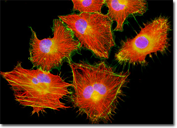
The baby hamster kidney (BHK-21) fibroblasts featured in the digital image above were resident in a culture immunofluorescently labeled for microtubules with primary anti-tubulin mouse monoclonal antibodies followed by goat anti-mouse Fab fragments conjugated to the cyanine dye, Cy3. In addition, the cells were labeled with Alexa Fluor 488 conjugated to phalloidin and DAPI, targeting the filamentous actin network and nuclear DNA, respectively. Images were recorded in grayscale with a QImaging Retiga Fast-EXi camera system coupled to an Olympus BX-51 microscope equipped with bandpass emission fluorescence filter optical blocks provided by Omega Optical. During the processing stage, individual image channels were pseudocolored with RGB values corresponding to each of the fluorophore emission spectral profiles.
Additional Fluorescence Images of Baby Hamster Kidney (BHK-21) Cells
Mitochondrial Localization by Fluorescent Proteins in Baby Hamster Kidney Fibroblasts - An adherent culture of baby hamster kidney fibroblast (BHK-21) cells was transfected with a pDsRed-Mitochondria plasmid subcellular localization vector, thus localizing a red fluorescent protein tag to the intracellular mitochondrial network. The culture was subsequently fixed, permeabilized and labeled with DAPI and Alexa Fluor 488 conjugated to phalloidin, targeting DNA and filamentous actin, respectively.
The Golgi Complex in BHK-21 Cell Cultures - A culture of baby hamster kidney cells was fixed, permeabilized, and blocked with 10-percent normal goat serum in phosphate-buffered saline prior to immunofluorescent labeling with rabbit primary antibodies to giantin, a protein resident in the Golgi complex of mammalian cells. The culture was subsequently stained with a mixture of goat anti-rabbit secondary antibody fragments (heavy and light chain) conjugated to Cy2. In addition, the culture was labeled for mitochondria with MitoTracker Red CMXRos, and for DNA with Hoechst 33342.
BACK TO THE CULTURED CELLS FLUORESCENCE GALLERY
BACK TO THE FLUORESCENCE GALLERY