
Fluorescence Digital Image Gallery
Human Cortical Neuronal Cells (HCN-1A Line)
The source for the HCN-1A cell line was cortical tissue removed from a patient undergoing hemispherectomy for intractable seizures. The patient was an 18-month-old female suffering from unilateral megalencephaly. Also known as hemimegalencephaly, this condition is characterized by the overgrowth of all or part of one of the cerebral hemispheres.
Though rare, unilateral megalencephaly is a very serious condition with a poor prognosis. Patients with this disorder often have a large, asymmetrical head and commonly experience severe mental retardation and untreatable seizures. Unilateral and other types of megalencephaly are generally believed to be related to a disturbance in the regulation of cell proliferation. Treatments for these conditions are symptomatic and supportive in nature.
HCN-1A cells stain positively for a number of neuronal markers including neuron specific enolase. They are also positive for tubulin, vimentin, somatostatin, glutamate, gamma aminobutyric acid, cholecystokinin-8, and vasoactive intestinal peptide. The cells are negative, however, for glial fibrillary acidic protein and myelin basis protein. HCN-1A cells can be induced to differentiate when cultured with a mixture of nerve growth factor, dibutyryl cyclic adenosine monophosphate, and 1-isobutyl-3-methylxanthine. Differentiation is accompanied by mature morphology and a deceleration of growth. Unlike HCN-2, a neuronal cell line derived from cortical tissue removed from a patient undergoing hemispherectomy for intractable seizures associated with Rasmussen's encephalitis, the growth rate of HCN-1A cells is not affected by phorbol esters. Both cell lines are, however, useful as models for the study of neuronal processes.
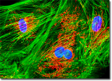
The culture of HCN-1A neuronal cells exhibited in the digital image above was labeled with MitoTracker Red CMXRos, Alexa Fluor 488 conjugated to phalloidin, and DAPI, targeting mitochondria, filamentous actin, and nuclear DNA, respectively. Images were recorded in grayscale with a QImaging Retiga Fast-EXi camera system coupled to an Olympus BX-51 microscope equipped with bandpass emission fluorescence filter optical blocks provided by Omega Optical. During the processing stage, individual image channels were pseudocolored with RGB values corresponding to each of the fluorophore emission spectral profiles.
Additional Fluorescence Images of Human Cortical Neuronal Cells
F-Actin Distribution in HCN-1A Neuronal Cells - The human cortical neuron culture (HCN-1A) featured in this section was immunofluorescently labeled with anti-tubulin mouse monoclonal primary antibodies followed by goat anti-mouse Fab fragments conjugated to Cy2. In addition, the culture was stained for the cytoskeletal F-actin network with Alexa Fluor 568 conjugated to phalloidin, and for DNA in the cell nucleus with Hoechst 33258.
BACK TO THE CULTURED CELLS FLUORESCENCE GALLERY
BACK TO THE FLUORESCENCE GALLERY