Fluorescence Digital Image Gallery
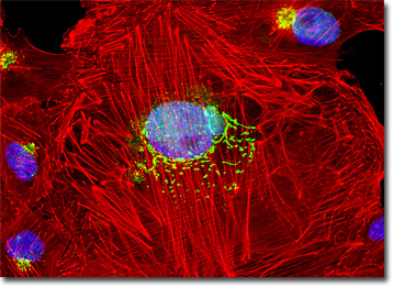
Normal African Green Monkey Kidney Epithelial Cells (Vero Line)
The Vero epithelial cell line was established in 1962 by Y. Yasumura and Y. Kawakita at the Chiba University in Chiba, Japan. The tissue from which the line was derived was obtained from the kidney of a healthy adult African green monkey. Although widely used in transfections and vaccine production, Vero cells are also often utilized in the detection of verotoxins, a group of interrelated toxins produced by some strains of Escherichia coli that are a key cause of hemorrhagic colitic and hemolytic uremic syndrome in humans.

The array of viruses that Vero cells are susceptible to is broad and includes polioviruses, simian virus 5 (SV5), simian virus 40 (SV40), rubeola, rubellavirus, reoviruses, simian adenoviruses, Getah, Ndumu, Pixuna, Ross River, Semliki Forest, Paramaribo, Kokobera, Modoc, Murutucu, Germiston, Guaroa, Pongola, and Tacaribe. The Vero cell line is negative, however, for reverse transcriptase and is resistant to Stratford, Apeu, Caraparu, Madrid, Nepuyo, and Ossa viruses.
In recent years, Vero cells have been under consideration for use in the development of new kinds of treatments for various diseases, such as diabetes. Characterized by the inability to metabolize glucose either from the destruction of the pancreatic beta cells that are responsible for producing insulin (Type 1) or a decreased ability to respond to insulin (Type 2), diabetes is of great concern in the medical community, especially since it is the leading cause of kidney disease. In 2002, a group of researchers working in Ireland reported that modified Vero cells show promise for cell therapy that could be used to treat Type 1, and possibly some cases of Type 2, diabetes. The approach suggested by the group involves engineering Vero cells to behave as beta cell substitutes. A potential advantage, noted by the group, of using such artificial beta substitutes rather than real beta cells from other sources is that non-beta cells may be able to subsist in the body unrecognized by the autoimmune response that characteristically attacks the cells in Type I diabetes patients.
The culture of normal African green monkey kidney epithelial cells (Vero line) presented in the digital image above was labeled with Oregon Green 488 conjugated to the lectin wheat germ agglutinin. The cells were also stained with Alexa Fluor 568 conjugated to phalloidin and DAPI, which target filamentous actin and nuclear DNA, respectively. Images were recorded in grayscale with a QImaging Retiga Fast-EXi camera system coupled to an Olympus BX-51 microscope equipped with bandpass emission fluorescence filter optical blocks provided by Omega Optical. During the processing stage, individual image channels were pseudocolored with RGB values corresponding to each of the fluorophore emission spectral profiles.
Additional Fluorescence Images of Normal African Green Monkey Kidney Epithelial Cells (Vero Line)
Vero African Green Monkey Kidney Cells with MitoTracker Red CMXRos, Alexa Fluor 488, and DAPI - One of the most useful fluorophore combinations for visualizing internal cellular details includes MitoTracker Red CMXRos to target mitochondria, Alexa Fluor 488 conjugated to phalloidin for the filamentous actin network, and DAPI to localize the nuclei. The cell culture illustrated in this section was stained with this protocol.
Immunofluorescence of the Microtubule Network in Normal African Green Monkey Kidney Cells - The microtubule network was imaged in a Vero cell culture by treating the fixed and permeabilized cells with mouse anti-alpha-tubulin primary antibodies, followed by goat anti-mouse secondary antibodies conjugated to Alexa Fluor 568. Nuclei were counterstained with SYTOX Green.
Visualizing Structural Features of the Golgi Complex and Nucleus in Vero Cell Cultures - In order to examine structural features of the Golgi complex and nucleus at relatively high magnification, a log-phase culture of normal African green monkey (Vero) cells was fixed, permeabilized, blocked with normal goat serum, and then treated with rabbit anti-giantin (Golgi protein) primary antibodies followed by goat anti-rabbit secondary antibodies (IgG) conjugated to Alexa Fluor 568. The nuclei were counterstained with Hoechst 33258.
Classical Staining Patterns in African Green Kidney (Vero Line) Epithelial Cells - The now traditional and popular combination of MitoTracker Red CMXRos, Alexa Fluor 488 conjugated to phalloidin, and Hoechst 33342 was used to triple-label an adherent culture of Vero African Green Monkey Kidney cells. Note the unusual filamentous actin network, and the large number of mitochondria surrounding the nuclei in these cells.
Epithelial Vero Cell Culture Labeled with Soybean Agglutinin - An adherent culture of African green monkey kidney epithelial cells was labeled with Alexa Fluor 488 conjugated to soybean agglutinin, a lectin isolated from Glycine max that selectively binds terminal alpha- and beta-N-acetylgalactosamine and galactopyranosyl residues. In addition, the cells were labeled with Alexa Fluor 568 conjugated to phalloidin and DAPI, targeting the cytoskeletal F-actin network and DNA, respectively.
Nuclear Pore Complex Proteins in African Green Monkey Kidney Epithelial Cells - A log phase culture of Vero cells was fixed, permeabilized, blocked with 10-percent normal goat serum, and then treated with mouse anti-NPCP (nuclear pore complex proteins) primary antibodies followed by secondary antibodies conjugated to Alexa Fluor 568. The secondary cocktail contained Alexa Fluor 488 conjugated to phalloidin to label the filamentous actin network, and the nuclei were counterstained with Hoechst 33342.
Visualizing the Tubulin and Actin Cytoskeletal Networks in Vero Cell Cultures - Elements of the cytoskeletal and microtubule networks were imaged in a Vero cell culture by treating the fixed and permeabilized cells with mouse anti-alpha-tubulin primary antibodies, followed by goat anti-mouse secondary antibodies conjugated to Alexa Fluor 568. Mixed with the secondary antibody cocktail was Alexa Fluor 350 conjugated to phalloidin to target filamentous actin. Nuclei were counterstained with SYTOX Green.
Normal African Green Monkey Kidney Cells with Alexa Fluor 488, Alexa Fluor 568, and Hoechst 33258 - A log phase culture of Vero cells was fixed, permeabilized, blocked, and treated with mouse anti-C-protein (myocardial) monoclonal primary antibodies followed by goat anti-mouse secondary antibodies conjugated to Alexa Fluor 568. Mixed in with the secondary reagent was Alexa Fluor 488 conjugated to phalloidin to label the filamentous actin cytoskeleton. Nuclei were counterstained with Hoechst 33258.
Distribution of Histones, Peroxisomes, and Filamentous Actin in Vero Cell Cultures - In a double immunofluorescence labeling experiment, an adherent culture of normal African green monkey kidney cells was treated with a cocktail of mouse anti-histones (pan) and rabbit anti-PMP 70 (peroxisomal membrane protein) primary antibodies, followed by goat anti-mouse and anti-rabbit secondary antibodies conjugated to Alexa Fluor 568 and Alexa Fluor 488, respectively, to target the nuclear histone proteins and peroxisomes. The filamentous actin network was imaged with Alexa Fluor 350 conjugated to phalloidin.
Targeting the Endoplasmic Reticulum in Normal African Green Monkey Cell Cultures with Concanavalin A - A monolayer culture of adherent Vero epithelial cells was stained with Alexa Fluor 568 conjugated to the lectin concanavalin A, which selectively binds to alpha-mannopyranosyl and alpha-glucopyranosyl residues (primarily in the endoplasmic reticulum). Alexa Fluor 488 conjugated to phalloidin and Hoechst 33258 were also used to label the culture, targeting filamentous actin and nuclear DNA, respectively.
Simultaneous Visualization of Clathrin Assemblies and the Golgi Complex in Vero Cells - In a double immunofluorescence experiment, an adherent monolayer culture of African green monkey kidney cells was fixed, permeabilized, blocked with normal goat serum, and treated with a cocktail of mouse anti-clathrin (heavy chain) and rabbit anti-giantin (Golgi) primary antibodies, followed by goat anti-mouse and anti-rabbit secondary antibodies (IgG) conjugated to Alexa Fluor 488 and Alexa Fluor 568, respectively. The nuclei were counterstained with Hoechst 33258.
BACK TO THE CULTURED CELLS FLUORESCENCE GALLERY
BACK TO THE FLUORESCENCE GALLERY
