
Fluorescence Digital Image Gallery
Pig Kidney Epithelial cells (LLC-PK1)
The LLC-PK1 cell line was derived from the kidney of a normal, healthy male pig (Sus scrofa) that was between 3 and 4 weeks of age. The pig was a member of the Hampshire breed, which was initially imported to America from Hampshire County, England in the early 1800s. Commonly utilized in laboratories around the world, the LLC-PK1 line exhibits typical epithelial morphology.
Cellular products of the LLC-PK1 porcine kidney line include plasminogen activator, a substance that stimulates fibrinolysis. In recent years, plasminogen activator has been included in drugs used in thrombosis therapies since it facilitates the dissolution of small blood clots. The cells also produce large amounts of cytokeratin. The LLC-PK1 line is often used as a model for epithelial tissue, as well as in a wide spectrum of pharmacologic and metabolic research investigations.
Part of the urinary system, a kidney, such as the one that served as the source for the LLC-PK1 cell line, is a major vertebrate excretory organ. The primary function of the organ is to separate urea, toxins, and other types of waste from the blood, while maintaining suitable water, salt, and electrolyte levels. The nephron, which consists of a renal corpuscle and a renal tubule, is the basic filtering unit of the kidney. A kidney belonging to an average human adult typically contains more than a million nephrons. Together they are capable of filtering the blood at an impressive rate, processing the entire five-quart water content of the human circulatory system about every 45 minutes. Over the course of a single day, therefore, each kidney filters approximately 160 quarts of material. However, only about 1 percent of this material is excreted, the rest being reabsorbed by the nephrons.
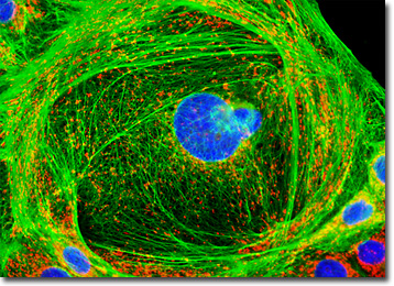
The adherent culture of LLC-PK1 epithelial cells featured in the digital image above was immunofluorescently labeled with primary anti-cytokeratin (pan) mouse monoclonal antibodies followed by goat anti-mouse Fab fragments conjugated to Alexa Fluor 488. In addition, the specimen was stained with MitoTracker Red CMXRos and DAPI, which bind with mitochondria and DNA, respectively. Images were recorded in grayscale with a QImaging Retiga Fast-EXi camera system coupled to an Olympus BX-51 microscope equipped with bandpass emission fluorescence filter optical blocks provided by Omega Optical. During the processing stage, individual image channels were pseudocolored with RGB values corresponding to each of the fluorophore emission spectral profiles.
Additional Fluorescence Images of Pig Kidney Epithelial (LLC-PK1) Cells
Cytokeratin Intermediate Filaments and the Golgi Network in LLC-PK1 Epithelial Cells - A log phase culture of pig kidney (LLC-PK1) cells was immunofluorescently labeled with primary anti-cytokeratin (pan) mouse monoclonal antibodies followed by goat anti-mouse Fab fragments conjugated to Oregon Green 488. Golgi bodies were simultaneously targeted with rabbit anti-giantin primary antibodies, followed by goat anti-rabbit secondaries conjugated to Texas Red. Nuclei were counterstained with Hoechst 33258.
Pig Kidney Epithelial Cells with MitoTracker Red CMXRos, SYTOX Green, and Marina Blue - In this section, the featured digital image presents a culture of pig kidney cells that was immunofluorescently labeled anti-cytokeratin (pan) mouse monoclonal primary antibodies followed by goat anti-mouse Fab fragments conjugated to Marina Blue. The cells were also labeled with MitoTracker Red CMXRos and SYTOX Green, targeting the intracellular mitochondrial network and DNA, respectively.
Double Immunofluorescence of Nuclear Pore Complex Proteins and Tight Junctions in LLC-PK1 Adherent Cell Cultures - Epithelial cell tight junctions and nuclear pore complex proteins were simultaneously imaged in normal pig kidney cells with a cocktail of mouse anti-NPCP and rabbit anti ZO-3 primary antibodies, followed by goat anti-mouse and anti-rabbit secondary antibodies conjugated to Alexa Fluor 488 and Alexa Fluor 568, respectively.
Microtubule Distribution in LLC-PK1 Cells - A culture of pig kidney epithelial (LLC-PK1) cells was immunofluorescently labeled with anti-tubulin mouse monoclonal primary antibodies followed by goat anti-mouse Fab fragments conjugated to Cy3, targeting the intracellular microtubule network. The culture was also stained for DNA with the ultraviolet-absorbing probe DAPI.
Pig Kidney Epithelial Cells with Alexa Fluor 568, Oregon Green 488, and DAPI - The adherent culture of LLC-PK1 cells presented in this section was stained for F-actin with Alexa Fluor 568 conjugated to phalloidin, and for DNA with 4',6-diamidino-2-phenylindole (DAPI). The cells were additionally labeled with Oregon Green 488 conjugated to lectin PNA, a protein derived from peanuts that binds to specific carbohydrate groups on proteins or cell membranes.
Sialic Acid Residue Distribution in LLC-PK1 Epithelial Cells - In order to target the cytoskeletal F-actin network and nuclei present in a LLC-PK1 monolayer culture, the cells were first stained with Alexa Fluor 488 conjugated to phalloidin and propidium iodide, respectively. The culture was also labeled with the probe Alexa Fluor 350 conjugated to wheat germ agglutinin, a lectin that selectively binds to sialic acid residues, which are found in both mucoproteins and glycoproteins.
Immunofluorescent Labeling of Cytokeratin in Pig Kidney Cell Cultures - The culture of pig kidney epithelial cells featured in this section was immunofluorescently labeled with anti-cytokeratin (pan) mouse monoclonal primary antibodies followed by goat anti-mouse Fab fragments conjugated to Pacific Blue. The specimen was subsequently stained with MitoTracker Red CMXRos and SYTOX Green to label the mitochondrial network and nuclear DNA, respectively.
LLC-PK1 Cells with Cy3, Alexa Fluor 488, and DAPI - A log phase culture of LLC-PK1 epithelial cells was immunofluorescently labeled with anti-tubulin mouse monoclonal primary antibodies followed by goat anti-mouse Fab fragments conjugated to Cy3, targeting the intracellular microtubular network. In addition, the cells were labeled for cytoskeletal F-actin with Alexa Fluor 488 conjugated to phalloidin, and for nuclear DNA with DAPI.
Traditional Fluorescence Staining Patterns in LLC-PK1 Cells - Adherent LLC-PK1 cells were treated with the traditional triple-fluorophore combination of MitoTracker Red CMXRos, Alexa Fluor 488 conjugated to phalloidin and Hoechst 33258. These probes label the mitochondria, filamentous actin network, and DNA in the nucleus, respectively.
Cytokeratin and Golgi Complex Distribution in Pig Kidney Epithelial Cell Cultures - Many epithelial cell cultures are good candidates for immunofluorescence labeling with cytokeratin. The LLC-PK1 culture featured in this section was treated with mouse anti-cytokeratin (pan) primary antibodies followed by goat anti-mouse secondaries conjugated to Texas Red. Simultaneously, the Golgi complex was visualized with rabbit anti-giantin primaries and goat anti-rabbit secondaries conjugated to Oregon Green 488. Nuclei were counterstained with Hoechst 33342.
Simultaneous Visualization of the Microtubule and Actin Cytoskeletal Networks in LLC-PK1 Cells - A healthy culture of pig kidney epithelial cells (LLC-PK1) was fixed, permeabilized, and treated with mouse anti-alpha-tubulin primary antibodies followed by a cocktail of goat anti-mouse Fab fragments conjugated to fluorescein and Alexa Fluor 568 conjugated to phalloidin. Nuclei were counterstained with DAPI.
Normal Pig Kidney Epithelial Cells with MitoTracker Red CMXRos, SYTOX Green, and Marina Blue - Log phase LLC-PK1 cells were bathed in growth medium containing MitoTracker Red CMXRos for one hour, and then fixed, permeabilized, and blocked with 10-percent normal goat serum. The culture was subsequently treated with mouse anti-cytokeratin (pan) primary antibodies followed by goat anti-mouse secondary antibodies conjugated to Marina Blue. DNA in the nuclei was visualized using SYTOX Green.
The Actin Cytoskeletal Network in Closely Packed LLC-PK1 Epithelial Cells - An adherent culture of closely packed pig kidney epithelial cells was immunofluorescently stained with mouse anti-alpha-tubulin primary antibodies followed by goat anti-mouse Fab fragments conjugated to the cyanine dye, Cy3. The filamentous actin network was revealed by including Alexa Fluor 488 conjugated to phalloidin with the secondary antibody solution, and the nuclei were subsequently counterstained with DAPI.
Targeting the Golgi Complex in Pig Kidney Epithelial Cells with Lectins - In this section, the featured culture of pig kidney (LLC-PK1) cells was labeled with Oregon Green conjugated to lectin GS-II, which is isolated from the seeds of a tropical legume and often utilized as a selective stain for the Golgi apparatus. The culture was additionally labeled for the cytoskeletal filamentous actin network with Alexa Fluor 568 conjugated to phalloidin, and for nuclear DNA with the ultraviolet-absorbing probe DAPI.
Normal Pig Kidney (LLC-PK1) Cells with Alexa Fluor 488, Alexa Fluor 568, and Hoechst 33258 - Simultaneous localization of tight junctions and the nuclear pore complex proteins (NPCP) was performed with a double immunofluorescence experiment with LLC-PK1 cells using mouse anti-NPCP and rabbit anti-ZO-3 primary antibodies. The subcellular targets were visualized using goat anti-mouse and anti-rabbit secondary antibodies (IgG) conjugated to Alexa Fluor 488 and Alexa Fluor 568, respectively. DNA in the nuclei was counterstained using Hoechst 33258.
Histone and Peroxisome Distribution in LLC-PK1 Cell Cultures - In a double immunofluorescence experiment, an adherent monolayer culture of normal pig kidney epithelial (LLC-PK1) cells was fixed, permeabilized, blocked with 10 percent normal goat serum, and treated with a cocktail of mouse anti-histones (pan) and rabbit anti-PMP 70 (peroxisomal membrane protein) primary antibodies, followed by goat anti-mouse and anti-rabbit secondary antibodies (IgG) conjugated to Texas Red and Alexa Fluor 488, respectively. The filamentous actin network was counterstained with Alexa Fluor 350 conjugated to phalloidin.
BACK TO THE CULTURED CELLS FLUORESCENCE GALLERY
BACK TO THE FLUORESCENCE GALLERY