
Fluorescence Digital Image Gallery
Mouse Embryo Teratocarcinoma Epithelial Cells (P19 Line)
The P19 cell line was derived from an embryonal carcinoma induced in a C3H/He mouse strain of the species Mus musculus. Similar to other embryonal carcinoma cells, cells of the P19 line are capable of differentiating into an array of cell types and are, therefore, described as pluripotent.
Though they can be maintained and propagated in tissue culture in an undifferentiated state, when exposed to 500 nM of retinoic acid, P19 cells can be induced to differentiate into neural and glial-like cells. In the presence of 0.5 to 1.0 percent dimethylsulfoxide (DMSO), however, the cells differentiate to form cardiac and skeletal muscle-like elements, but do not form neural or glial-like cells. When introduced simultaneously to both DMSO and retinoic acid, P19 cells differentiate as if they were exposed only to retinoic acid.
The specific type of embryonal carcinoma from which the P19 cell line was established is a teratocarcinoma, a malignant germ cell tumor. Teratocarcinomas most frequently occur in the male testes, but may also affect female ovaries and other organs of the body. Some of the tumors include a number of well-differentiated cells, which can lead to the development of tissues in the tumor that are very different from normal tissues found in that area. For instance, ovarian teratocarcinomas have been found that have grown hair and teeth. Typically, teratocarcinomas of the testes are less differentiated than other types and are, therefore, characteristically the most common variety to serve as sources for embryonal carcinoma cells intended for research purposes.
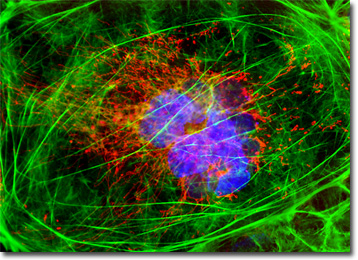
The mouse embryo teratocarcinoma (P19) cell culture presented in the digital image above was labeled for mitochondria and the cytoskeletal filamentous actin network with MitoTracker Red CMXRos and Alexa Fluor 488 conjugated to phalloidin, respectively. In addition, the cells were counterstained for DNA with the ultraviolet-absorbing probe DAPI. Images were recorded in grayscale with a QImaging Retiga Fast-EXi camera system coupled to an Olympus BX-51 microscope equipped with bandpass emission fluorescence filter optical blocks provided by Omega Optical. During the processing stage, individual image channels were pseudocolored with RGB values corresponding to each of the fluorophore emission spectral profiles.
BACK TO THE CULTURED CELLS FLUORESCENCE GALLERY
BACK TO THE FLUORESCENCE GALLERY