
Fluorescence Digital Image Gallery
Female Rat Kangaroo Kidney Epithelial Cells (PtK1 Line)
The PtK1 cell line was derived in 1962 from the kidney of a normal adult female Potorous tridactylus, a species of rat kangaroo prevalent in Australia. The epithelial line is believed to be the first permanent cell line of marsupial origin to be established. PtK1 cells are positive for keratin by immunoperoxidase and fluorescence staining and are susceptible to vesicular stomatitis (Indiana strain).
PtK1 epithelial cells are resistant, however, to poliovirus 2 and are also negative for reverse transcriptase, indicating the lack of integral retrovirus genomes. The cell line has been primarily utilized in mitotic research because of the low number, large size, and distinct morphology of the chromosomes of the species from which it was established.
Chromosomes are the microscopic, threadlike strands of a cell that carry genes, hereditary units that are primarily comprised of DNA. In eukaryotes, chromosomes are found in a membrane-bound nucleus and contain RNA as well as DNA. All species of organisms exhibit a characteristic chromosome number. The karyotype of a human, for example, contains 46 chromosomes, while that of a female Potorous tridactylus, such as the one that served as the source of the PtK1 cell line, typically only contains 11. Due to this relatively small number, studying chromosomes of the PtK1 line is often considered a simpler endeavor than examining those of other cell lines. The line is especially well suited to be an observational model for mitosis, since the cells tend to stay relatively flat even when dividing. For similar reasons, the PtK2 cell line, which was derived from the kidney of a normal adult male rat kangaroo, is also widely utilized in the study of mitosis and chromosome function, structure, and evolution.
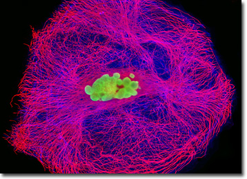
In order to simultaneously visualize both the actin and tubulin networks in PtK1 cells to capture the digital image presented above, a fixed and permeabilized culture was treated with mouse anti-alpha-tubulin primary antibodies, followed by a cocktail of goat anti-mouse secondary antibodies conjugated to Alexa Fluor 568 mixed together with Alexa Fluor 350 conjugated to phalloidin. Nuclei were counterstained with SYTOX Green. Images were recorded in grayscale with a QImaging Retiga Fast-EXi camera system coupled to an Olympus BX-51 microscope equipped with bandpass emission fluorescence filter optical blocks provided by Omega Optical. During the processing stage, individual image channels were pseudocolored with RGB values corresponding to each of the fluorophore emission spectral profiles.
BACK TO THE CULTURED CELLS FLUORESCENCE GALLERY
BACK TO THE FLUORESCENCE GALLERY