
Fluorescence Digital Image Gallery
Bovine Pulmonary Artery Endothelial Cells (BPAE Line)
The BPAE cell line was initiated in January 1979 by P. Del Vecchio from the main stem of a pulmonary artery belonging to a young cow (Bos taurus). Pulmonary arteries, which extend from the heart to the lungs, are the only arteries in the mammalian body that carry dark, unoxygenated blood.
The BPAE line of endothelial cells is positive for bovine diarrhea virus, one of the most important known bovine viral pathogens, which causes a broad array of clinical syndromes that result in significant losses in the beef industry each year. BPAE cells are also positive for angiotensin converting enzyme (ACE), an enzyme that is intricately involved in the maintenance of blood pressure and volume. Due to this fact, BPAE cells are often utilized in hypertension research as well as studies of atherosclerosis and coronary heart disease.
Endothelial cells, such as those of the BPAE line, are generally flat and elongate in shape. When found in the body lining the pulmonary arteries, these cells are usually arranged so that their long axis is oriented parallel to the direction of blood flow in the vessel. The cells are connected to one another via both gap junctions and tight junctions. An organized array of membrane channels, a gap junction allows adjacent cells to communicate through direct passage of small molecules and ions. In contrast, tight junctions act as diffusion barriers that restrict the dispersal of molecules to adjacent cells with varying degrees of success. Tight junctions are found at the most apical point between neighboring endothelial cells and form a band around the entire circumference of the cell.
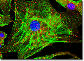
The adherent culture of BPAE cells featured in the digital image above was immunofluorescently labeled with anti-vinculin mouse monoclonal primary antibodies followed by goat anti-mouse Fab heavy and light chain fragments conjugated to Rhodamine Red-X. In addition, the specimen was simultaneously stained for DNA with the ultraviolet-absorbing probe DAPI, and for the cytoskeletal filamentous actin network with Alexa Fluor 488 conjugated to phalloidin. Images were recorded in grayscale with a QImaging Retiga Fast-EXi camera system coupled to an Olympus BX-51 microscope equipped with bandpass emission fluorescence filter optical blocks provided by Omega Optical. During the processing stage, individual image channels were pseudocolored with RGB values corresponding to each of the fluorophore emission spectral profiles.
Additional Fluorescence Images of Bovine Pulmonary Artery (BPAE) Cells
Distribution of Mitochondria in BPAE Cell Cultures - A culture of bovine pulmonary artery endothelial cells was stained with MitoTracker Red CMXRos, vividly labeling the intracellular mitochondrial network. The specimen was also labeled for filamentous actin and DNA with Alexa Fluor 488 (green emission) conjugated to phalloidin and DAPI (blue emission), respectively.
BACK TO THE CULTURED CELLS FLUORESCENCE GALLERY
BACK TO THE FLUORESCENCE GALLERY