Fluorescence Digital Image Gallery
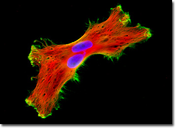
Human Brain Glioma Cells (U-118 MG Line)
The U-118 MG cell line is one of several cell lines derived from malignant gliomas by J. Ponten and associates in the late 1960s. The source for the cells of this particular line was a 50-year-old Caucasian male. The morphology of U-118 MG line is mixed and both glioblastoma and astrocytoma cells are present.

U-118 MG cells are very similar to the glioblastoma U-138 MG cell line, though the line is supposed to have been derived from a different source. Studies have found that both of these lines have identical VNTR (variable number of tandem repeats) patterns and similar STR (short tandem repeat) patterns in DNA analysis experiments. They also have in common at least six derivative marker chromosomes. Mycoplasma contamination was detected and eliminated from the U-118 MG cell line in 1987 through treatment of cultures with BM-cycline over a six-week period. The cells have been demonstrated to be tumorigenic in nude mice subcutaneously inoculated.
A glioma, such as the one that served as the basis of the U-118 MG cell line, is a malignant brain tumor. As suggested by their name, gliomas originate from glial cells, which comprise the tissue that surrounds and supports neurons in the central nervous system. Although all brain tumors are extremely serious, gliomas may be particularly dangerous because they tend to grow quickly and readily spread within the brain. Approximately 20,000 victims in the United States are diagnosed with glioma each year, more than half of which die in less than two years. The chance of survival for a particular individual, however, depends upon many factors, including age, stage of tumor advancement, and the area of the brain affected.
The isolated U-118 MG glioma cell presented in the digital image above was resident in a culture that was immunofluorescently labeled with anti-tubulin mouse monoclonal primary antibodies followed by goat anti-mouse Fab fragments conjugated to Texas Red. In addition, the specimen was stained with Alexa Fluor 488 conjugated to phalloidin and Hoechst 33342, targeting the cytoskeletal filamentous actin network and DNA in the cell nucleus, respectively. Images were recorded in grayscale with a QImaging Retiga Fast-EXi camera system coupled to an Olympus BX-51 microscope equipped with bandpass emission fluorescence filter optical blocks provided by Omega Optical. During the processing stage, individual image channels were pseudocolored with RGB values corresponding to each of the fluorophore emission spectral profiles.
Additional Fluorescence Images of Human Brain Glioma (U-118 MG) Cells
Histone and Peroxisome Distribution in Human Brain Glioma (U-118 MG) Cell Cultures - In a double immunofluorescence experiment, an adherent monolayer culture of U-118 MG cells was fixed, permeabilized, blocked with 10 percent normal goat serum, and treated with a cocktail of mouse anti-histones (pan) and rabbit anti-PMP 70 (peroxisomal membrane protein) primary antibodies, followed by goat anti-mouse and anti-rabbit secondary antibodies (IgG) conjugated to Texas Red and Alexa Fluor 488, respectively. The filamentous actin network was counterstained with Alexa Fluor 350 conjugated to phalloidin.
BACK TO THE CULTURED CELLS FLUORESCENCE GALLERY
BACK TO THE FLUORESCENCE GALLERY
