
Fluorescence Digital Image Gallery
Rat Kidney Mesangial Cells (RMC Line)
The RMC cell line was derived from the kidney tissue of a 3-month-old male rat (Rattus norvegicus) belonging to the Sprague-Dawley strain. The mesangial cells were immortalized at passage eight with the plasmid pSV3-Neo, which codes simian virus 40 (SV40) large T-antigen, and are maintained in the presence of the antibiotic, G-418.
The rat kidney cells express normal genes of the wild type mesangial cells and grow adherently to glass and polymer surfaces in monolayer culture. RMC cells are positive for desmin and vimentin, but are negative for cytokeratin 8. Mesangial cells are specialized cells usually associated with glomeruli that are crucial for kidney function. Thus, the RMC line and other lines of mesangial cells are widely utilized in kidney research.
Two different groups of mesangial cells have been identified in the kidneys. Intraglomerular mesangial cells are located within the renal corpuscle and have been demonstrated to be phagocytic. These cells are generally believed to primarily function in the removal of debris from the glomerular basement membrane (GBM) that is the principal component of the filtration barrier of a glomerulus. In clinical studies it has been found that intraglomerular mesangial cells flourish in patients with certain renal diseases in which atypically large amounts of protein and protein complexes are caught in the GBM. Moreover, since mesangial cells are contractile, it is also possible that they function in some manner in regulating the flow of blood through glomeruli. When the specialized cells appear outside of the renal corpuscle along the vascular pole they are referred to as extraglomerular mesangial cells. These cells comprise part of the juxtaglomerular apparatus, which apparently functions in sensing and regulating blood composition and volume.
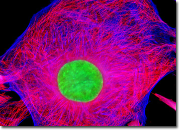
In order to visualize the microtubules present in the rat kidney mesangial cells illustrated in the digital image above, an adherent RMC culture was immunofluorescently labeled with anti-tubulin mouse monoclonal primary antibodies followed by goat anti-mouse Fab fragments conjugated to Alexa Fluor 568. In addition, the cells were labeled for the cytoskeletal filamentous actin network with Alexa Fluor 350 conjugated to phalloidin, and for cell nuclei with the classic nucleic acid stain, SYTOX Green. Images were recorded in grayscale with a QImaging Retiga Fast-EXi camera system coupled to an Olympus BX-51 microscope equipped with bandpass emission fluorescence filter optical blocks provided by Omega Optical. During the processing stage, individual image channels were pseudocolored with RGB values corresponding to each of the fluorophore emission spectral profiles.
Additional Fluorescence Images of Rat Kidney Mesangial (RMC) Cells
Rat Kidney Mesangial Cells with MitoTracker Red CMXRos, Alexa Fluor 488, and Hoechst 33258 - Using the popular triple fluorophore combination of MitoTracker Red CMXRos, Alexa Fluor 488 conjugated to phalloidin, and Hoechst 33258, the adherent culture of rat kidney mesangial cells featured in this section was stained to visualize the mitochondria, filamentous actin network, and nuclei.
BACK TO THE CULTURED CELLS FLUORESCENCE GALLERY
BACK TO THE FLUORESCENCE GALLERY