
Fluorescence Digital Image Gallery
Human Skin Epidermoid Carcinoma Epithelial Cells (A-431 Line)
The A-431 cell line, which was derived from an epidermoid carcinoma excised from the skin tissue of an 85-year-old female, is one of a succession of cell lines established from solid tumors by a research team led by D. J. Giard. The epithelial line is tumorigenic, forming rapidly growing subcutaneous tumors in immunosuppressed mice and colonies in soft agar. Also known as cutaneous squamous cell carcinoma, epidermoid carcinoma of the skin is a malignant tumor of epidermal keratinocytes, a condition that has increased in frequency considerably over the last century, perhaps due in part to the ongoing depletion of the protective ozone layer.
Epidermoid carcinoma is one of the most common types of malignant tumors that affect the integumentary system, accounting for approximately 20 percent of all skin cancers. A variety of causes may lead to the condition, such as exposure to arsenic, coal, tar, paraffin, or the human papilloma virus, but the vast majority of cases involve overexposure to ultraviolet light, typically from the harsh rays of the sun, but also from tanning beds and other sources of the radiation. Those at greatest risk for developing cutaneous epidermoid carcinoma include people with light eyes and a fair complexion that tends to burn rather than tan, inhabitants of areas where the sun is notoriously bright throughout the year, such as Arizona and Florida, those that work outdoors, and the elderly or others who may possess a weakened immune system.
Epidermoid carcinoma of the skin often appears as a raised reddish or pink bump, which may be waxy and smooth or scaly and rough. The tumor may also occur as a lesion that tends to bleed easily or crust over and is most commonly found in areas that chronically encounter the sun (forehead, ears, etc.). The prognosis for those that develop this form cancer is typically quite positive since the tumor tends to remain localized for a more extensive period of time than many other varieties of the disease. Thus, surgery is often sufficient to eliminate the tumor, though it may not necessarily prohibit the cancer from recurring. Also, as with other cancers, when metastasis does occur, treatment becomes much difficult and may involve a multiplicity of therapies, such as radiation and chemotherapy, rather than solely surgery.
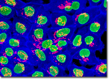
The A-431 cells illustrated above were grown to mid-log phase in adherent monolayer culture, and then fixed, permeabilized, and treated with a cocktail of mouse anti-nuclear pore complex protein NPCP and rabbit anti-giantin (Golgi apparatus) primary antibodies, followed by goat anti-mouse and anti-goat secondary antibodies conjugated to Oregon Green 488 and Alexa Fluor 568, respectively. The filamentous actin cytoskeletal network was simultaneously imaged using Alexa Fluor 350 conjugated to phalloidin. Images were recorded in grayscale with a QImaging Retiga Fast-EXi camera system coupled to an Olympus BX-51 microscope equipped with bandpass emission fluorescence filter optical blocks provided by Omega Optical. During the processing stage, individual image channels were pseudocolored with RGB values corresponding to each of the fluorophore emission spectral profiles.
BACK TO THE CULTURED CELLS FLUORESCENCE GALLERY
BACK TO THE FLUORESCENCE GALLERY