
Introduction to Cell and Virus Structure
At first glance, the petal of a flower or the skin on the back of a human hand may seem smooth and seamless, as if they were composed of a single, indistinct substance. In reality, however, many tiny individual units called cells make up these objects and almost all other components of plants and animals. The average human body contains over 75 trillion cells, but many life forms exist as single cells that perform all the functions necessary for independent existence. Most cells are far too small to be seen with the naked eye and require the use of high-power optical and electron microscopes for careful examination.
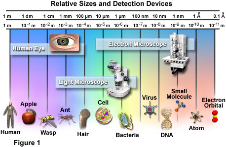
The relative scale of biological organisms as well as the useful range of several different detection devices are illustrated in Figure 1. The most basic image sensor, the eye, was the only means humans had of visually observing the world around them for thousands of years. Though excellent for viewing a wide variety of objects, the power of the eye has its limits, anything smaller than the width of a single human hair being able to pass unnoticed by the organ. Therefore, when light microscopes of sufficient magnifying capability were developed in the late 1600s, a whole new world of tiny wonders was discovered. Electron microscopes, invented in the mid-twentieth century, made it possible to detect even tinier objects than light microscopes, including smaller molecules, viruses, and DNA. The detection power of most electron microscopes used today, however, stops just short of being able to visualize such incredibly small structures as the electron orbital systems of individual atoms. Atoms are considered the smallest units of an element that have the characteristics of that element, but cells are the smallest structural units of an organism capable of functioning independently.
Yet, until the mid-seventeenth century, scientists were unaware that cells even existed. It wasn't until 1665 that biologist Robert Hooke observed through his microscope that plant tissues were divided into tiny compartments, which he termed "cellulae" or cells. It took another 175 years, however, before scientists began to understand the true importance of cells. In their studies of plant and animal cells during the early nineteenth century, German botanist Matthias Jakob Schleiden and German zoologist Theodor Schwann recognized the fundamental similarities between the two cell types. In 1839, they proposed that all living things are made up of cells, the theory that gave rise to modern biology.
Since that time, biologists have learned a great deal about the cell and its parts; what it is made of, how it functions, how it grows, and how it reproduces. The lingering question that is still being actively investigated is how cells evolved, i.e., how living cells originated from nonliving chemicals.
Numerous scientific disciplines�physics, geology, chemistry, and evolutionary biology�are being used to explore the question of cellular evolution. One theory speculates that substances vented into the air by volcanic eruptions were bombarded by lightning and ultraviolet radiation, producing larger, more stable molecules such as amino acids and nucleic acids. Rain carried these molecules to the Earth's surface where they formed a primordial soup of cellular building blocks.
A second theory proposes that cellular building blocks were formed in deep-water hydrothermal vents rather than in puddles or lakes on the Earth's surface. A third theory speculates that these key chemicals fell to earth on meteorites from outer space.
Given the basic building blocks and the right conditions, it would seem to be just a matter of time before cells begin to form. In the laboratory, lipid (fat) molecules have been observed joining together to produce spheres that are similar to a cell's plasma membrane. Over millions of years, perhaps it is inevitable that random collisions of lipid spheres with simple nucleic acids, such as RNA, would result in the first primitive cells capable of self-replication.
For all that has been learned about cells in over 300 years, hardly the least of which is the discovery of genetic inheritance and DNA, cell biology is still an exciting field of investigation. One recent addition is the study of how physical forces within the cell interact to form a stable biomechanical architecture. This is called "tensegrity" (a contraction of "tensional integrity"), a concept and word originally coined by Buckminster Fuller. The word refers to structures that are mechanically stable because stresses are distributed and balanced throughout the entire structure, not because the individual components have great strength.
In the realm of living cells, tensegrity is helping to explain how cells withstand physical stresses, how they are affected by the movements of organelles, and how a change in the cytoskeleton initiates biochemical reactions or even influences the action of genes. Some day, tensegrity may even explain the mechanical rules that caused molecules to assemble themselves into the first cells.
Animal Cells - Animal cells are typical of the eukaryotic cell type, enclosed by a plasma membrane and containing a membrane-bound nucleus and organelles.
Bacteria - One of the earliest prokaryotic cells to have evolved, bacteria have been around for at least 3.5 billion years and live in almost every imaginable environment.
Plant Cells - The basic plant cell has a similar construction to the animal cell, but does not have centrioles, lysosomes, cilia, or flagella. It does have additional structures, including a rigid cell wall, central vacuole, plasmodesmata, and chloroplasts.
Virus Structure - Viruses are not alive in the strict sense of the word, but reproduce and have an intimate, if parasitic, relationship with all living organisms.
Cells in Motion - In multicellular tissues, such as those found in animals and humans, individual cells employ a variety of locomotion mechanisms to maneuver through spaces in the extracellular matrix and over the surfaces of other cells. Examples are the rapid movement of cells in developing embryos, organ-to-organ spreading of malignant cancer cells, and the migration of neural axons to synaptic targets. Unlike single-celled swimming organisms, crawling cells in culture do not possess cilia or flagella, but tend to move by coordinated projection of the cytoplasm in repeating cycles of extension and retraction that deform the entire cell. The digital videos presented in this gallery investigate animal cell motility patterns in a wide variety of morphologically different specimens.
Fluorescence Microscopy of Cells in Culture - Serious attempts at the culture of whole tissues and isolated cells were first undertaken in the early 1900s as a technique for investigating the behavior of animal cells in an isolated and highly controlled environment. The term tissue culture arose because most of the early cells were derived from primary tissue explants, a technique that dominated the field for over 50 years. As established cell lines emerged, the application of well-defined normal and transformed cells in biomedical investigations has become an important staple in the development of cellular and molecular biology. This fluorescence image gallery explores over 30 of the most common cell lines, labeled with a variety of fluorophores using both traditional staining methods as well as immunofluorescence techniques.
Observing Mitosis with Fluorescence Microscopy - Mitosis, a phenomenon observed in all higher eukaryotes, is the mechanism that allows the nuclei of cells to split and provide each daughter cell with a complete set of chromosomes during cellular division. This, coupled with cytokinesis (division of the cytoplasm), occurs in all multicellular plants and animals to permit growth of the organism. Digital imaging with fluorescence microscopy is becoming a powerful tool to assist scientists in understanding the complex process of mitosis on both a structural and functional level.
Mitosis Java Tutorial - Explore the stages of mitosis in eukaryotic cells with this interactive Java tutorial. Step through prophase, metaphase, anaphase, and telophase as the chromosomes slowly condense, align, and divide before being segregated into daughter cells.
Cell Digestion and the Secretory Pathway - The primary sites of intracellular digestion are organelles known as the lysosomes, which are membrane-bounded compartments containing a variety of hydrolytic enzymes. Lysosomes maintain an internal acidic environment through the use of a hydrogen ion pump in the lysosomal membrane that drives ions from the cytoplasm into the lumenal space of the organelles. The high internal acidity is necessary for the enzymes contained in lysosomes to exhibit their optimum activity. Hence, if the integrity of a lysosomal membrane is compromised and the enzymatic contents are leaked into the cell, little damage is done due to the neutral pH of the cytoplasm. If numerous lysosomes rupture simultaneously, however, the cumulative action of their enzymes can result in autodigestion and the death of the cell.
BACK TO MOLECULAR EXPRESSIONS HOME