
Virus Structure
Viruses are not plants, animals, or bacteria, but they are the quintessential parasites of the living kingdoms. Although they may seem like living organisms because of their prodigious reproductive abilities, viruses are not living organisms in the strict sense of the word.
Without a host cell, viruses cannot carry out their life-sustaining functions or reproduce. They cannot synthesize proteins, because they lack ribosomes and must use the ribosomes of their host cells to translate viral messenger RNA into viral proteins. Viruses cannot generate or store energy in the form of adenosine triphosphate (ATP), but have to derive their energy, and all other metabolic functions, from the host cell. They also parasitize the cell for basic building materials, such as amino acids, nucleotides, and lipids (fats). Although viruses have been speculated as being a form of protolife, their inability to survive without living organisms makes it highly unlikely that they preceded cellular life during the Earth's early evolution. Some scientists speculate that viruses started as rogue segments of genetic code that adapted to a parasitic existence.
All viruses contain nucleic acid, either DNA or RNA (but not both), and a protein coat, which encases the nucleic acid. Some viruses are also enclosed by an envelope of fat and protein molecules. In its infective form, outside the cell, a virus particle is called a virion. Each virion contains at least one unique protein synthesized by specific genes in its nucleic acid. Viroids (meaning "viruslike") are disease-causing organisms that contain only nucleic acid and have no structural proteins. Other viruslike particles called prions are composed primarily of a protein tightly integrated with a small nucleic acid molecule.
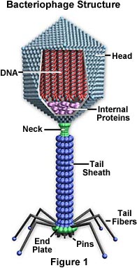
Viruses are generally classified by the organisms they infect, animals, plants, or bacteria. Since viruses cannot penetrate plant cell walls, virtually all plant viruses are transmitted by insects or other organisms that feed on plants. Certain bacterial viruses, such as the T4 bacteriophage, have evolved an elaborate process of infection. The virus has a "tail" which it attaches to the bacterium surface by means of proteinaceous "pins." The tail contracts and the tail plug penetrates the cell wall and underlying membrane, injecting the viral nucleic acids into the cell. Viruses are further classified into families and genera based on three structural considerations: 1) the type and size of their nucleic acid, 2) the size and shape of the capsid, and 3) whether they have a lipid envelope surrounding the nucleocapsid (the capsid enclosed nucleic acid).
There are predominantly two kinds of shapes found amongst viruses: rods, or filaments, and spheres. The rod shape is due to the linear array of the nucleic acid and the protein subunits making up the capsid. The sphere shape is actually a 20-sided polygon (icosahedron).
The nature of viruses wasn't understood until the twentieth century, but their effects had been observed for centuries. British physician Edward Jenner even discovered the principle of inoculation in the late eighteenth century, after he observed that people who contracted the mild cowpox disease were generally immune to the deadlier smallpox disease. By the late nineteenth century, scientists knew that some agent was causing a disease of tobacco plants, but would not grow on an artificial medium (like bacteria) and was too small to be seen through a light microscope. Advances in live cell culture and microscopy in the twentieth century eventually allowed scientists to identify viruses. Advances in genetics dramatically improved the identification process.
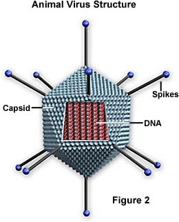
Capsid - The capsid is the protein shell that encloses the nucleic acid; with its enclosed nucleic acid, it is called the nucleocapsid. This shell is composed of protein organized in subunits known as capsomers. They are closely associated with the nucleic acid and reflect its configuration, either a rod-shaped helix or a polygon-shaped sphere. The capsid has three functions: 1) it protects the nucleic acid from digestion by enzymes, 2) contains special sites on its surface that allow the virion to attach to a host cell, and 3) provides proteins that enable the virion to penetrate the host cell membrane and, in some cases, to inject the infectious nucleic acid into the cell's cytoplasm. Under the right conditions, viral RNA in a liquid suspension of protein molecules will self-assemble a capsid to become a functional and infectious virus.
Envelope - Many types of virus have a glycoprotein envelope surrounding the nucleocapsid. The envelope is composed of two lipid layers interspersed with protein molecules (lipoprotein bilayer) and may contain material from the membrane of a host cell as well as that of viral origin. The virus obtains the lipid molecules from the cell membrane during the viral budding process. However, the virus replaces the proteins in the cell membrane with its own proteins, creating a hybrid structure of cell-derived lipids and virus-derived proteins. Many viruses also develop spikes made of glycoprotein on their envelopes that help them to attach to specific cell surfaces.
Nucleic Acid - Just as in cells, the nucleic acid of each virus encodes the genetic information for the synthesis of all proteins. While the double-stranded DNA is responsible for this in prokaryotic and eukaryotic cells, only a few groups of viruses use DNA. Most viruses maintain all their genetic information with the single-stranded RNA. There are two types of RNA-based viruses. In most, the genomic RNA is termed a plus strand because it acts as messenger RNA for direct synthesis (translation) of viral protein. A few, however, have negative strands of RNA. In these cases, the virion has an enzyme, called RNA-dependent RNA polymerase (transcriptase), which must first catalyze the production of complementary messenger RNA from the virion genomic RNA before viral protein synthesis can occur.
The Influenza (Flu) Virus - Next to the common cold, influenza or "the flu" is perhaps the most familiar respiratory infection in the world. In the United States alone, approximately 25 to 50 million people contract influenza each year. The symptoms of the flu are similar to those of the common cold, but tend to be more severe. Fever, headache, fatigue, muscle weakness and pain, sore throat, dry cough, and a runny or stuffy nose are common and may develop rapidly. Gastrointestinal symptoms associated with influenza are sometimes experienced by children, but for most adults, illnesses that manifest in diarrhea, nausea, and vomiting are not caused by the influenza virus though they are often inaccurately referred to as the "stomach flu." A number of complications, such as the onset of bronchitis and pneumonia, can also occur in association with influenza and are especially common among the elderly, young children, and anyone with a suppressed immune system.
The Human Immunodeficiency Virus (HIV) - The virus responsible for HIV was first isolated in 1983 by Robert Gallo of the United States and French scientist Luc Montagnier. Since that time, a tremendous amount of research focusing upon the causative agent of AIDS has been carried out and much has been learned about the structure of the virus and its typical course of action. HIV is one of a group of atypical viruses called retroviruses that maintain their genetic information in the form of ribonucleic acid (RNA). Through the use of an enzyme known as reverse transcriptase, HIV and other retroviruses are capable of producing deoxyribonucleic acid (DNA) from RNA, whereas most cells carry out the opposite process, transcribing the genetic material of DNA into RNA. The activity of the enzyme enables the genetic information of HIV to become integrated permanently into the genome (chromosomes) of a host cell.