
Fluorescence Microscopy
Practical Aspects of Fluorescence Filter Combinations
The terminology applied to fluorescence filters has become a jumble as a result of various initials utilized by different manufacturers to identify their filters. In this section, we attempt to make some order of this confusing terminology and specifically address how the various filter combinations can be utilized with specific fluorophores. Basically there are three categories of filters to be sorted out: exciter filters, barrier filters and dichromatic beamsplitters (dichroic mirrors), which are usually combined to produce a filter cube or block similar to the one illustrated in Figure 1. Proper selection of filters is the key to successful fluorescence microscopy.
The following sections contain links to review articles and interactive Java tutorials describing a variety of fluorescence filter combinations from the major manufacturers. Additional information is provided with regards to illumination excitation balancers, selection of fluorophores, and matching specific probes with filter combinations.
Brief Overview of Fluorescence Filters - Microscope manufacturers provide proprietary filter combinations (often referred to as cubes or blocks) that contain a combination of dichroic mirrors and filters capable of exciting fluorescent chromophores and diverting the resulting secondary fluorescence to the eyepieces or camera tube. A wide spectrum of filter cubes is available from most major manufacturers, which now produce filter sets capable of imaging most of the common chromophores in use today.
Basic Aspects of Light Filters - Many filters work by absorbing light, while others reflect unwanted light, but pass a selected region of wavelengths. The color temperature of light can be fine-tuned with filters to produce a spectrum of light having the characteristics of bright daylight, the evening sky, indoor tungsten illumination, or some variation in between. Filters are useful for adjusting the contrast of colored regions as they are represented in black and white photography or to add special effects in color photography. Specialized dichroic filters can be used to polarize light, while heat-absorbing filters can limit infrared wavelengths (and heat), allowing only visible light to pass through. Harmful ultraviolet rays can be exclusively removed from visible light by filters, or the intensity of all wavelengths (ultraviolet, visible, and infrared) can be reduced to specific ranges by neutral density filters. The most sophisticated filters operate by the principles of interference and can be adjusted to pass narrow bands (or even a single wavelength) of light while reflecting all others in a specific direction.
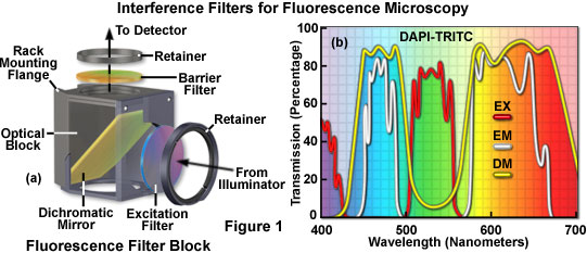
Interference Filters for Fluorescence Microscopy - The performance of high-resolution fluorescence microscopy imaging systems and related quantitative applications, especially as applied in living cell and tissue studies, requires precise optimization of fluorescence excitation and detection strategies. Fluorescence microscopy techniques could not have advanced so dramatically in recent years without significant developments in every dimension of the current state of the art, including the optical microscopes, the biology and chemistry of fluorophores, and perhaps most important, filter technology. The utilization of highly specialized and advanced thin film interference filters has enhanced the versatility and scope of fluorescence techniques, far beyond the capabilities afforded by the earlier use of gelatin and glass filters relying on the absorption properties of embedded dyes.
Nikon Fluorescence Filter Combinations - Epi-Fluorescence interference filter combinations are housed in a filter cube and include an excitation filter, dichroic mirror (or beamsplitter), and a barrier (or emission) filter. Use this guide in selecting the appropriate filter set to match the spectral excitation and emission characteristics of chromophores used in fluorescence microscopy experiments. Although the filter combinations discussed in this section are marketed by Nikon, the spectral profile and fluorophore response characteristics also apply to filters from other major microscope and aftermarket filter manufacturers.
Acousto-Optic Tunable Filters (AOTFs) - Several benefits of the AOTF combine to greatly enhance the versatility of the latest generation of confocal instruments, and these devices are becoming increasing popular for control of excitation wavelength ranges and intensity. The primary characteristic that facilitates nearly every advantage of the AOTF is its capability to allow the microscopist control of the intensity and/or illumination wavelength on a pixel-by-pixel basis while maintaining a high scan rate. This single feature translates into a wide variety of useful analytical microscopy tools, which are even further enhanced in flexibility when laser illumination is employed.
Introduction to Fluorophores - Widefield fluorescence and laser scanning confocal microscopy rely heavily on secondary fluorescence emission as an imaging mode, primarily due to the high degree of sensitivity afforded by the techniques coupled with the ability to specifically target structural components and dynamic processes in chemically fixed as well as living cells and tissues. Many fluorescent probes are constructed around synthetic aromatic organic chemicals designed to bind with a biological macromolecule (for example, a protein or nucleic acid) or to localize within a specific structural region, such as the cytoskeleton, mitochondria, Golgi apparatus, endoplasmic reticulum, and nucleus. Other probes are employed to monitor dynamic processes and localized environmental variables, including concentrations of inorganic metallic ions, pH, reactive oxygen species, and membrane potential. Fluorescent dyes are also useful in monitoring cellular integrity (live versus dead and apoptosis), endocytosis, exocytosis, membrane fluidity, protein trafficking, signal transduction, and enzymatic activity. In addition, fluorescent probes have been widely applied to genetic mapping and chromosome analysis in the field of molecular genetics.
Spectral Bleed-Through Artifacts in Confocal Microscopy - The spectral bleed-through of fluorescence emission (often termed crossover or crosstalk), which occurs due to the very broad bandwidths and asymmetrical spectral profiles exhibited by many of the common fluorophores, is a fundamental problem that must be addressed in both widefield and laser scanning confocal fluorescence microscopy. The phenomenon is usually manifested by the emission of one fluorophore being detected in the photomultiplier channel or through the filter combination reserved for a second fluorophore. Bleed-through artifacts often complicate the interpretation of experimental results, particularly if subcellular colocalization of fluorophores is under investigation or quantitative measurements are necessary, such as in resonance energy transfer (FRET) and photobleaching (FRAP) studies.
Interactive Java Tutorials
Interference Filters - Recent technological achievements in bandpass filter design have led to the relatively inexpensive construction of thin-film interference filters featuring major improvements in wavelength selection and transmission performance. These filters operate by transmitting a selected wavelength region with high efficiency while rejecting, through reflection and destructive interference, all other wavelengths. Explore how interference filters operate by selectively transmitting constructively reinforced wavelengths while simultaneously eliminating unwanted light with this interactive tutorial.
Matching Fluorescent Probes with Fluorescence Filter Blocks - Modern fluorescence microscope instrumentation utilizes a combination of filters in conjunction with a dichromatic beam splitter to satisfy the excitation and emission requirements of the fluorescent probe(s) used to label the specimen. When these components are chosen appropriately, the microscope provides an essential mechanism for selective excitation of specimen fluorophores, and the subsequent isolation of much weaker fluorescence emission necessary for image formation. By carefully matching excitation and emission filter properties with the function of the dichromatic beamsplitter, labeled specimen features are imaged on a dark background with maximum sensitivity. This interactive tutorial enables visitors to determine optimum choices among current Nikon fluorescence filter blocks for maximizing the efficiency of excitation and emission with specific fluorescent probes.
Matching Fluorescence Filter Blocks with Fluorescent Probes - The essential feature of any fluorescence microscope is to provide a mechanism for excitation of the specimen with selectively filtered illumination followed by isolation of the much weaker fluorescence emission using a second filter to enable image formation on a dark background with maximum sensitivity. These conditions are satisfied in modern fluorescence instruments by a combination of filters that coordinate excitation and emission requirements based on the action and properties of the dichromatic beamsplitter. This interactive tutorial enables visitors to determine the optimum fluorophore characteristics necessary to maximize the efficiency of excitation and emission in conjunction with current Olympus fluorescence filter blocks.
Balancing Arc-Discharge Lamp Excitation Illumination - Fine-tuning of the fluorescence microscope excitation spectrum for imaging dual or multiply labeled specimens can be readily accomplished with a split-filter excitation balancer, which contains tandem shortpass and longpass interference filters that are translated across the illumination aperture to adjust the arc-discharge lamp wavelength distribution profile. This interactive tutorial explores how the Nikon Eclipse i-Series excitation balancer system affects the fluorescence emission intensity of multiply labeled specimens when employed in conjunction with dual and triple excitation band filter combinations.
Fluorescence Filter Noise Terminator - An innovative new fluorescence filter block design by Nikon helps to eliminate the possibility of residual stray light that occurs in the microscope fluorescence optical pathway, vastly improving the emission signal-to-noise ratio. Termed the Noise Terminator, this technology directs deviated stray light away from the objective light collection path, resulting in a dramatic improvement in image contrast. This interactive tutorial demonstrates how the Noise Terminator technology functions.
Fluorescent Probe Excitation Efficiency - The absorption and fluorescence emission spectral profiles of a fluorophore are two of the most important criteria that must be scrutinized when selecting probes for applications in laser scanning confocal microscopy. In addition to the wavelength range of the absorption and emission bands, the molar extinction coefficient for absorption and the quantum yield for fluorescence emission should be considered. At laser excitation levels that do not saturate the fluorophore, fluorescence intensity is directly proportional to the product of the extinction coefficient and the quantum yield. This interactive tutorial examines how this relationship can be utilized to match fluorophores with specific lasers for confocal microscopy.
Acousto-Optic Tunable Filters - Wavelength selection is of fundamental importance in many arenas of the optical sciences, including fluorescence spectroscopy and microscopy. Electro-optic devices, such as the acousto-optic tunable filter (AOTF), are increasingly being employed to modulate the wavelength and amplitude of illuminating laser light in the latest generation of confocal microscopes. These filters do not suffer from the mechanical constraints, speed limitations, image shift, and vibration associated with rotating filter wheels, and can easily accommodate several laser systems tuned to different output wavelengths. In addition, acousto-optic filters do not deteriorate when exposed to heat and intense light as do fluorescence interference filters.
Liquid Crystal Tunable Filters - Liquid crystal tunable filters (LCTFs) use electrically controlled liquid crystal elements to select a specific visible wavelength of light for transmission through the filter at the exclusion of all others. This type of filter is ideal for use with electronic imaging devices, such as charge-coupled devices (CCDs), because it offers excellent imaging quality with a simple linear optical pathway.
Fluorochrome and Fluorescence Filter Combination Data Tables
In this section, we have compiled a listing of data on the most popular fluorochromes including excitation and emission characteristics, suggested cube utilization, and application data. This information can be accessed through the links provided below.
Fluorochrome Data Table - As a guide to fluorophores for confocal and widefield fluorescence microscopy, the table presented in this section lists many commonly-used fluorochromes, with their respective peak absorption and emission wavelengths and suggested laser illumination sources. While the authors assume responsibility for the accurate reporting of the data as published in various reliable sources, several caveats must be given. Fluorochrome dyes are environmentally sensitive and different results will be obtained with different solvents and applications. In reviewing the literature, one will frequently find somewhat different data supplied for the identical fluorochrome. There are also, in many instances, several sub-varieties of a fluorochrome.
Ex/Em/Cubes/Applications - A comprehensive table of excitation and emission spectral maxima, and excitation suggestions, listed by application.
Excitation and Emission Data - Excitation and emission maximum wavelengths listed alphabetically by fluorochrome name.
Olympus Fluorescence Vertical Illuminator Cubes - Data tables of the Olympus cubes for older models, including the U-URA, BH-2, Vanox-3, and IMT-3 fluorescent illuminators.
Fluorochromes and Suitable Cubes - A compilation of spectral characteristics of major fluorochromes as well as sample fluorescence photomicrographs are also included in a short to long wavelength sequence.
Olympus UIS Fluorescence Mirror Units - Designed to present the technical specifications of UIS fluorescence mirror units, this brochure offers a detailed unit selection chart based on fluorochrome characteristics. Mirror unit descriptions, a graphic representation of the characteristics of mirror unit wavelength, as well as information on custom fluorescence mirror units, fluorescence excitation balancers, and light source spectral characteristics are also provided.
Contributing Authors
Mortimer Abramowitz - Olympus America, Inc., Two Corporate Center Drive., Melville, New York, 11747.
Christopher Hardee, Roy Kinoshita, Travis Wakefield, and Robert Johnson - Omega Optical, Inc., 210 Main Street, Brattleboro, Vermont, 05301.
Turan Erdogan - Semrock, Inc., 3625 Buffalo Road, Rochester, New York, 14624.
Douglas B. Murphy - Department of Cell Biology and Anatomy and Microscope Facility, Johns Hopkins University School of Medicine, 725 N. Wolfe Street, 107 WBSB, Baltimore, Maryland 21205.
Kenneth R. Spring - Scientific Consultant, Lusby, Maryland, 20657.
Matthew J. Parry-Hill, Thomas J. Fellers, and Michael W. Davidson - National High Magnetic Field Laboratory, 1800 East Paul Dirac Dr., The Florida State University, Tallahassee, Florida, 32310.
BACK TO FLUORESCENCE MICROSCOPY