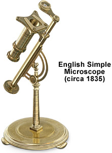
English Simple Brass Microscope
This microscope was presented to the Royal Society by Edward Milles Nelson in June of 1903. It is part of the general collection of the Royal Microscopical Society in London and is described by Gerard Turner in his book The Great Age of the Microscope.

The limb is triangular in shape and attaches to the solid brass pillar via a screw joint mechanism. Concentric with the joint is a slotted semicircular plate having an inner guide that controls the horizontal position (inclination) of the microscope body with a locking screw. Three bun feet placed at 120-degree spacings support the circular base, to which the pillar and microscope assembly are attached.
The microscope is not equipped with a coarse adjustment, but fine focus can be achieved via a knurled knob located at the base of the triangular rod that runs inside the limb. A simple convex condensing lens is positioned inside a brass sliding tube and is fixed to the bottom of the specimen stage. Specimens are secured to the stage with a pair of clips. A single objective is mounted in a brass housing that is fastened to the top of the limb with a small screw. The lens has a focal length of approximately one inch and incorporates a Lieberkuhn reflector. The entire microscope is fashioned from brass stock and displays a considerable degree of machinery marks that were not finished or polished.
BACK TO NINETEENTH CENTURY MICROSCOPES
