Fluorescence Digital Image Gallery
Female Rat Kangaroo Kidney Epithelial Cells (PtK1)
|
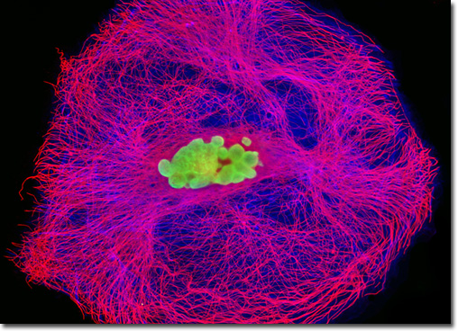
A number of nucleic acid stains have been developed for use with fluorescence microscopy, including the popular SYTOX line of cyanine dye derivatives. SYTOX stains are impermeant to live cells and are, therefore, often used as convenient indicators of living and dead cells within a culture. SYTOX Green is a member of the line that is especially well suited for use with both gram-positive and gram-negative bacteria, which necessitate an extremely bright signal. The dye also performs well as a DNA counterstain in combination with other probes in multicolor fluorescence microscopy applications. When bound to DNA, the fluorescence quantum yield of SYTOX Green is radically enhanced and in this bound state, the SYTOX Green/DNA complex exhibits an absorption maximum of 504 nanometers and an emission maximum of 523 nanometers. In order to simultaneously visualize both the actin and tubulin networks in PtK1 cells to capture the digital image presented above, a fixed and permeabilized culture was treated with mouse anti-alpha-tubulin primary antibodies, followed by a cocktail of goat anti-mouse secondary antibodies conjugated to Alexa Fluor 568 mixed together with Alexa Fluor 350 conjugated to phalloidin. Nuclei were counterstained with SYTOX Green. Images were recorded in grayscale with a QImaging Retiga Fast-EXi camera system coupled to an Olympus BX-51 microscope equipped with bandpass emission fluorescence filter optical blocks provided by Omega Optical. During the processing stage, individual image channels were pseudocolored with RGB values corresponding to each of the fluorophore emission spectral profiles. |
© 1995-2025 by Michael W. Davidson and The Florida State University. All Rights Reserved. No images, graphics, software, scripts, or applets may be reproduced or used in any manner without permission from the copyright holders. Use of this website means you agree to all of the Legal Terms and Conditions set forth by the owners.
This website is maintained by our
|