Fluorescence Digital Image Gallery
Baby Hamster Kidney Fibroblast Cells (BHK-21)
|
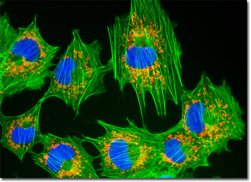
The red fluorescent protein DsRed was derived from the oral disk of a coral species belonging to the genus Discosoma. The absorption maximum of wild type DsRed is approximately 558 nanometers and the emission peak of the fluorescent protein is approximately 583 nanometers, in the yellow region of the visible spectrum. Some studies indicate that these maxima may be shifted to higher values through various mechanisms, such as the mutation of the lysine residue at position 83 to methionine. Mutagenesis has also been utilized to create DsRed variants with other useful characteristics, such as faster color maturation and increased solubility. Wild type DsRed and its variants are somewhat resistant to photobleaching and the effects of changes in pH. The fluorescent proteins are of most interest as expressible fluorescent reporter systems and conjugates that are complementary to green fluorescent protein (GFP). The adherent culture of baby hamster kidney fibroblast cells (BHK-21 line) featured in the digital image above was transfected with a pDsRed-Mitochondria plasmid subcellular localization vector, thus localizing a red fluorescent protein tag to the intracellular mitochondrial network. The culture was subsequently fixed and labeled with DAPI and Alexa Fluor 488 conjugated to phalloidin, targeting DNA and filamentous actin, respectively. Images were recorded in grayscale with a QImaging Retiga Fast-EXi camera system coupled to an Olympus BX-51 microscope equipped with bandpass emission fluorescence filter optical blocks provided by Omega Optical. During the processing stage, individual image channels were pseudocolored with RGB values corresponding to each of the fluorophore emission spectral profiles. |
© 1995-2025 by Michael W. Davidson and The Florida State University. All Rights Reserved. No images, graphics, software, scripts, or applets may be reproduced or used in any manner without permission from the copyright holders. Use of this website means you agree to all of the Legal Terms and Conditions set forth by the owners.
This website is maintained by our
|