
Observing Mitosis with Fluorescence Microscopy
Telophase
In telophase, the daughter chromosomes arrive at the spindle poles and are eventually redistributed into bulk chromatin. Individual chromosomes begin to decondense back into chromatin at this stage and start to become less clearly defined. Polymerized microtubular networks that formed the mitotic spindle during metaphase and anaphase are redistributed into cytoskeletal components, and RNA synthesis commences once again in the nucleus. The process of cytokinesis, where the cytoplasm is divided by cleavage into two daughter cells, also starts sometime in late anaphase and continues through telophase.
View a second, third, and fourth fluorescence image of telophase.
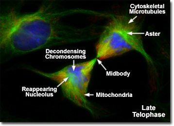
Presented in the digital fluorescence micrograph above is a pair of rat kangaroo (PtK2) kidney epithelial cells in the late stages of telophase. The chromatin is stained with a blue fluorescent probe (DAPI), while the cytoskeletal microtubule network and mitotic spindle are stained green (Alexa Fluor 488). Cellular mitochondria, which have been almost equally divided between newly formed daughter cells, are stained with a red dye (MitoTracker Red CMXRos). Note that the mitochondria are becoming interspersed throughout the cytoplasm, the previously condensed chromosomes are forming interphase chromatin, and the mitotic spindle is being redistributed into a cytoskeletal network. A thin bridge between the daughter cells, termed the midbody, contains remnants of polar microtubules from the mitotic spindle and is visible under the microscope for several hours after telophase has been completed.
After complete separation of the chromosomes and their extrusion to the spindle poles, the nuclear membrane begins to reform around each group of chromosomes at the opposite ends of the parent cell. The nucleoli also reappear in what will eventually become the two new daughter cell nuclei. When telophase is complete and the new cell membrane (or cell wall in the case of the higher plants) is being formed, the nuclei have almost matured to the pre-mitotic state. The final steps in telophase involve the initiation of plasma membrane cleavage between each of the new daughter cells to ultimately yield two separate cells during the next phase of cell division, known as cytokinesis.
During the telophase stage, the chromosomes uncoil and revert to their extended form, which is manifested in the homogeneous appearance of chromatin displayed by interphase chromosomes. Also in late telophase, the chromosomal fibers disappear and nucleoli develop at the nucleolar organizing sites. Nuclear envelopes start to form around the nuclei from remnants of the parent nuclear envelope and portions of the endomembrane system contained in each of the daughter cells. In addition, the cytoplasmic network of microtubules begins to once again define the cellular structure.
The nuclear membrane is reassembled during late telophase from vesicles that surround individual chromosomes, which eventually fuse into larger groups that finally associate to complete the structure. During this process, nuclear pores are established and the lamins, which have been dephosphorylated during anaphase, condense to reform the nuclear lamina. Upon complete reconstitution of the nuclear envelope assembly, the nuclear pores act as a gateway for nuclear proteins and the chromosomes begin to reform chromatin, an event that is probably triggered by the dephosphorylation of histone H1. At this point, RNA synthesis resumes and the nucleolus reappears.
Late in telophase, new centrioles are formed adjacent to existing copies of the organelles so that each daughter cell contains a complementary centriole pair. The cell membrane begins to constrict in a cleavage furrow between the separated chromosome sets, and the two daughter cells are ultimately connected only by the midbody (see the micrograph illustrated above), a bundle of residual spindle polar microtubules surrounded by a thin ribbon of cytoplasm. At the conclusion of telophase, mitosis is almost completed and the genetic information contained in the parent cell is equally distributed to each daughter. Following telophase, the cells become completely separated through a process known as cytokinesis or cleavage in animal cells.
BACK TO MITOSIS WITH FLUORESCENCE MICROSCOPY