Observing Mitosis with Fluorescence Microscopy
Telophase
|
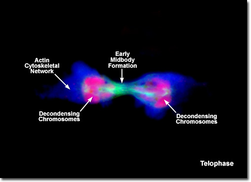
Presented in the digital fluorescence image above is a rat kangaroo (PtK2) cell in telophase. The chromosomes are stained with a red probe (Alexa Fluor 568), while the mitotic apparatus is green (Alexa Fluor 488). Actin filaments in the cytoskeleton are stained blue (Alexa Fluor 350). After complete separation of the chromosomes and their extrusion to the spindle poles, the nuclear membrane begins to reform around each group of chromosomes at the opposite ends of the parent cell. The nucleoli also reappear in what will eventually become the two new daughter cell nuclei. When telophase is complete and the new cell membrane (or cell wall in the case of the higher plants) is being formed, the nuclei have almost matured to the pre-mitotic state. The final steps in telophase involve the initiation of plasma membrane cleavage between each of the new daughter cells to ultimately yield two separate cells during the next phase of cell division, known as cytokinesis. |
© 1995-2025 by Michael W. Davidson and The Florida State University. All Rights Reserved. No images, graphics, software, scripts, or applets may be reproduced or used in any manner without permission from the copyright holders. Use of this website means you agree to all of the Legal Terms and Conditions set forth by the owners.
This website is maintained by our
|
