|
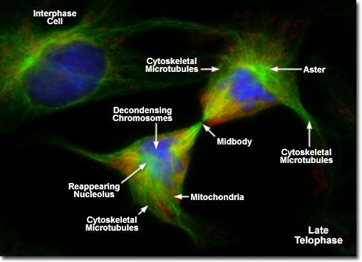
Presented in the digital fluorescence micrograph above is a pair of rat kangaroo (PtK2) kidney epithelial cells in the late stages of telophase. The chromatin is stained with a blue fluorescent probe (DAPI), while the cytoskeletal microtubule network and mitotic spindle are stained green (Alexa Fluor 488). Cellular mitochondria, which have been almost equally divided between newly formed daughter cells, are stained with a red dye (MitoTracker Red CMXRos). Note that the mitochondria are becoming interspersed throughout the cytoplasm, the previously condensed chromosomes are forming interphase chromatin, and the mitotic spindle is being redistributed into a cytoskeletal network. A thin bridge between the daughter cells, termed the midbody, contains remnants of polar microtubules from the mitotic spindle and is visible under the microscope for several hours after telophase has been completed.
|
