
Observing Mitosis with Fluorescence Microscopy
Anaphase
Almost immediately after the metaphase chromosomes are aligned at the metaphase plate, the two chromatids from each chromosome are pulled apart by the mitotic apparatus and migrate to the opposite spindle poles in a process known as anaphase. The kinetochore microtubules shorten as the chromatids are pulled toward opposite poles, while the polar microtubules subsequently elongate to assist in the separation. Anaphase typically is a rapid process that lasts only a few minutes, making it the shortest stage in mitosis.
View a second, third, and fourth fluorescence image of anaphase.
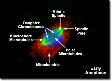
Presented in the digital fluorescence micrograph illustrated above is a single rat kangaroo (PtK2) kidney epithelial cell in the early stages of anaphase. The chromatin is stained with a blue fluorescent probe (DAPI), while the microtubule network (mitotic spindle) is stained green (Alexa Fluor 488) and cellular mitochondria are stained with a red dye (MitoTracker Red CMXRos). The paired centromeres from each chromosome have just begun to separate and the sister chromatids are being pulled apart by the kinetochore microtubules as they are shortened by the loss of tubulin monomers. Migration of the chromatids to opposite poles of the mitotic spindle occurs at a rate of approximately one micrometer per second in most animal cells.
During anaphase, the spatial position of the centromere with respect to the chromosome arms becomes obvious when imaged with the microscope. Because the sister chromatids are drawn towards the spindle poles by microtubules attached to the centromere, the arms appear to be trailing behind. Morphologically, the chromosomes can be divided into three classes. Metacentric chromosomes have the centromere located at or near the middle of the structure, resulting in two arms of equivalent (or nearly so) length. These chromosomes appear V-shaped when observed during anaphase. In contrast, a telocentric chromosome has the centromere positioned very near one end and appears to migrate as a single arm. A majority of the chromosomes, however, are acrocentric, with the centromere positioned somewhere between the center and the end of the chromatid to yield an L-shaped structure in the microscope.
The onset of anaphase is triggered by specific biochemical changes that occur to regulate the division cycle through modulation of the enzyme cascade responsible for cell-wide protein phosphorylation. Deactivation of the mitosis-inducing protein kinase (abbreviated MPF from the older term maturation-promoting factor) activity by proteolysis of cyclin subunits serves to initiate anaphase. Scientists speculate that the sister chromatids, which are held together along their length by a network of proteins, undergo a biochemical cleavage reaction that enables them to separate and begin the journey towards the spindle poles.
Anaphase is characterized by two distinct processes to separate the sister chromatids and move them to opposite spindle poles in preparation for cell division. The first process, termed anaphase A (or early anaphase), occurs with the shortening of the kinetochore microtubules to translocate the chromatids away from the metaphase plate and towards the spindle poles. Anaphase B (or late anaphase) is manifested by a further separation of the spindle poles brought about by lengthening of the polar microtubule network. The overall contribution of each process to the final separation of the sister chromatids varies depending upon the organism being studied. In animal cells, anaphase B begins soon after the chromatids split and terminates when the spindle has grown to approximately twice the metaphase length. In contrast, the cells of many lower eukaryotes exhibit a much larger separation distance during late anaphase, which can result in a spindle length increase up to 15 times the metaphase size.
When the chromosomes have completely migrated to the spindle poles, the kinetochore microtubules begin to disappear, although the polar microtubules continue to elongate. This is the junction between late anaphase and early telophase, the last stage in chromosome division. By the end of anaphase, each spindle pole has acquired an equivalent set of daughter chromosomes. The process of cytokinesis (splitting of the daughter cells) also begins during late anaphase with the initial formation of a contractile ring consisting of actin and myosin-II filaments positioned beneath the plasma membrane parallel to the metaphase plate. Later in the division cycle, the contractile ring will slowly converge to pull the membrane inward in order to divide the cell.
BACK TO MITOSIS WITH FLUORESCENCE MICROSCOPY