Observing Mitosis with Fluorescence Microscopy
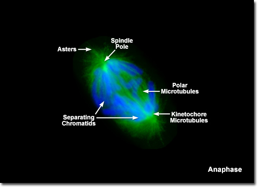
Anaphase
|
During anaphase, the spatial position of the centromere with respect to the chromosome arms becomes obvious when imaged with the microscope. Because the sister chromatids are drawn towards the spindle poles by microtubules attached to the centromere, the arms appear to be trailing behind. Morphologically, the chromosomes can be divided into three classes. Metacentric chromosomes have the centromere located at or near the middle of the structure, resulting in two arms of equivalent (or nearly so) length. These chromosomes appear V-shaped when observed during anaphase. In contrast, a telocentric chromosome has the centromere positioned very near one end and appears to migrate as a single arm. A majority of the chromosomes, however, are acrocentric, with the centromere positioned somewhere between the center and the end of the chromatid to yield an L-shaped structure in the microscope. |
© 1995-2025 by Michael W. Davidson and The Florida State University. All Rights Reserved. No images, graphics, software, scripts, or applets may be reproduced or used in any manner without permission from the copyright holders. Use of this website means you agree to all of the Legal Terms and Conditions set forth by the owners.
This website is maintained by our
|