Observing Mitosis with Fluorescence Microscopy
Anaphase
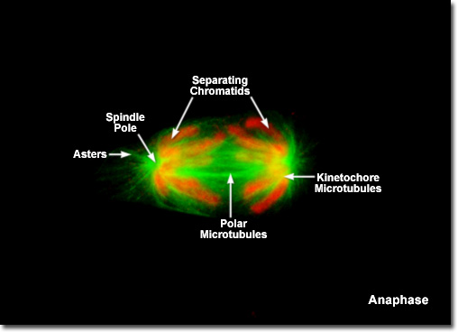
|
When the chromosomes have completely migrated to the spindle poles, the kinetochore microtubules begin to disappear, although the polar microtubules continue to elongate. This is the junction between late anaphase and early telophase, the last stage in chromosome division. By the end of anaphase, each spindle pole has acquired an equivalent set of daughter chromosomes. The process of cytokinesis (splitting of the daughter cells) also begins during late anaphase with the initial formation of a contractile ring consisting of actin and myosin-II filaments positioned beneath the plasma membrane parallel to the metaphase plate. Later in the division cycle, the contractile ring will slowly converge to pull the membrane inward in order to divide the cell. |
© 1995-2025 by Michael W. Davidson and The Florida State University. All Rights Reserved. No images, graphics, software, scripts, or applets may be reproduced or used in any manner without permission from the copyright holders. Use of this website means you agree to all of the Legal Terms and Conditions set forth by the owners.
This website is maintained by our
|