
Rheinberg Illumination
Rheinberg illumination, a form of optical staining, was initially demonstrated by the British microscopist Julius Rheinberg to the Royal Microscopical Society and the Quekett Club (England) over a hundred years ago. This technique is a striking variation of low to medium power darkfield illumination using colored gelatin or glass filters to provide rich color to both the specimen and background.
The Rheinberg technique can be compared to the more familiar darkfield illumination. In darkfield microscopy, the substage condenser is arranged so that the rays of light from the lamp, coming through the condenser, will pass through the specimen only at very oblique angles. The central area of the cone of light traversing the condenser is occluded by means of an opaque stop, large enough in diameter so as to prevent light from directly entering the microscope objective. In more sophisticated darkfield condensers--paraboloid, cardioid, Cassegrain, or Leuchtbild--the occlusion of the direct light and the utilization of oblique rays only are achieved by use of specially designed mirror surfaces.
| Interactive Tutorial | |||||||||||
|
|||||||||||
In darkfield illumination, the objective's numerical aperture is chosen (or reduced via an objective iris or funnel stop) so that light from the condenser cannot enter the objective directly. The only light that does enter the objective is light reflected, refracted, or diffracted by the specimen when the oblique rays from the condenser "strike" the specimen. The specimen then appears bright on an otherwise black field; hence the name darkfield illumination. The stark contrast of a bright object on a black field increases the visibility of already-resolved detail. A darkfield condenser can be approximated with the illustration in Figure 1 when the green central filter is substituted with an opaque stop, and the red oblique illumination filter is removed, allowing unfiltered white light to pass through.
In Rheinberg illumination (illustrated in Figure 1), which is related to darkfield, several kinds and shapes of filters are used. The oblique or outer light rays coming through a wide-open bright-field condenser pass through an annular (doughnut-shaped) filter of one or more colors (shown as the red filter in Figure 1 and the annular filters in Figure 2); the central rays of light pass through another spot-shaped filter fitting into the circular opening of the annular-shaped filter as shown for several combinations in Figure 2. The objective is used at full aperture. In this particular case, the specimen would appear as either red on a green background (Figure 2(a)), yellow on a blue background (Figure 2(b)), or green on a red background (Figure 2(c)).
The Rheinberg filters are prepared, or manufactured, in the form of transparent, annular colored rings with a circular opening in the center as depicted in Figure 3. In commercial versions of these filters, an accompanying set of transparent colored spots or "central filters" is made to fit snugly into the openings of the annular rings (Figure 4). The diameter of the central filter can be varied (to modulate background illumination intensity) by increasing the size of the narrow black rings that separate the filter from the outer ring (Figure 4(a-c)). Effective combinations include: red ring, violet central filter; yellow-orange ring, blue central filter, and so forth. These combinations can be explored using our Rheinberg Illumination Tutorial to adjust both the annular ring and central filter colors. Other visually effective images are produced by using a clear uncolored ring with a colored central filter. For example, a clear ring with a red central filter will produce white or colorless images on a red background.
In effect, the outer ring "becomes" the color of the specimen and the central filter "becomes" the color of the background. The outer ring can also be divided into alternating sectors of color as illustrated in Figure 5. The sector filters are especially effective in the study of warp-proof fabrics, crystal faces, diatoms, and wood sections where the length-width dimensions are displayed in contrasting colors.
For years, some microscope manufacturers sold Rheinberg filters in colored gels (similar to Kodak Wratten filters) that were precut to fit the substage filter ring of their microscopes. The annular rings had outside diameters of 31-35 millimeters and central filters usually had a diameter of 15-18 millimeters for use with a 10x - 0.25 NA objective. These filters can also be readily fabricated in the laboratory using either Kodak Wratten filters or colored filters that are widely available at scientific supply houses and optical component distributors. Gelatin filters can be prepared by cutting the central filter with a cork-borer of the proper diameter, and the annular rings can be prepared by carefully drawing a circle on the filter paper and cutting it out with a pair of scissors. In our laboratory, the support machinists have constructed a brass die that cuts annular filter circles that are the same diameter as our Rheinberg filter holder. This die can quickly stamp very smooth circles from sheets of acetate or gelatin colored filters. The center of the annular filter can be cut with the same cork-borer used to prepare the central filter.
| Interactive Tutorial | |||||||||||
|
|||||||||||
Sometimes, it is difficult to provide a solid housing for the central filter within the annular filter when these are both made of very thin acetate or gelatin filters. In this case, you can just tape the central filter over the center of the annular filter with double-sided tape, but remember that the central filter and annular filter colors will add, so use a very dark filter for the central filter. The central filter, in general, should be much darker than the annular filter to allow the specimen highlights to be in sharp contrast to the background. We often place two or three pieces of the same color central filters in a stack to adjust the transmittance of light through the central filter. Rheinberg filters can also be made using glass filters and this is very convenient when the substage condenser is fitted with a housing for these filters. In this case, it is best to simply tape the central filter onto the center of the glass filter. For those interested in making their own filters, a more complete discussion is presented in Needham's Practical Use of the Microscope, listed in our bibliography.
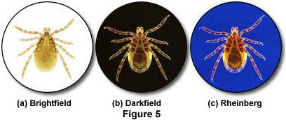
We have compared brightfield, darkfield, and Rheinberg illumination techniques using photomicrographs of a Deer tick (Ixodes dammini) in Figure 6. The first photomicrograph (a) illustrates the tick under brightfield illumination. The image is lacking in contrast and many details are hard to resolve. Figure 6(b) shows the same tick under darkfield illumination, where more contrast and detail are present and many of the features on the tick are apparent. Rheinberg illumination (Figure 6(c)) of the tick using a blue central filter and a yellow annular ring (see Figure 2(b)) cause an increased contrast effect, similar to darkfield, but with a pleasant blue background. In this example, the darkfield and Rheinberg illumination techniques yield remarkably better photomicrographs than does brightfield.
During the late 1930s, Carl Zeiss manufactured a special condenser, the Mikropolychromar, designed to produce beautiful Rheinberg images. This condenser is long out of production and is now virtually unobtainable, but it consisted of an aplanatic condenser under which there were three separately controlled diaphragms. The outermost diaphragm controlled the diameter of the field, and two smaller diaphragms controlled the light passing through the central disk. Annular transparent rings of various colors were part of the set. Accompanying these was a set of transparent glass central disks which fitted neatly into the central opening of the annular ring. This ingenious condenser has been used by one of the authors (M. Abramowitz) to produce striking photomicrographs at various magnifications, some of which have appeared in the publications Omni, Time-Life, Scientific American, and National Wildlife.
Rheinberg illumination is suitable for objectives ranging from 2x to 100x. However, in order to clearly separate the inner and outer colors, opaque metal or paper rings should be placed around the central filter. For example, the opaque rings might have an outside diameter of 15-18 millimeters for a 10x objective, and an outside diameter of 22 millimeters might be best for a 60x oil immersion objective. It is a good idea to experiment using central filters like the ones illustrated in Figure 4 to achieve the best results with Rheinberg illumination.
For studying fibers, protozoa, textiles, insects, wood sections, crystal, or other unstained low-contrast subjects, microscopists should consider adding Rheinberg illumination to their library of contrast techniques. It will be possible to view and photograph specimens yielding images that are visually enhanced, as well as aesthetically beautiful.
One final note from the authors: It is strongly suggested to the microscope manufacturers and/or independent accessory manufacturers that they produce Rheinberg condensers and filters for use by microscopists. The necessary components would be very inexpensive to produce and would greatly aid microscopists in setting up their microscopes for Rheinberg Illumination.
Contributing Authors
Mortimer Abramowitz - Olympus America, Inc., Two Corporate Center Drive., Melville, New York, 11747.
Michael W. Davidson - National High Magnetic Field Laboratory, 1800 East Paul Dirac Dr., The Florida State University, Tallahassee, Florida, 32310.