Fluorescence Digital Image Gallery
Human Brain Glioma Cells (U-118 MG)
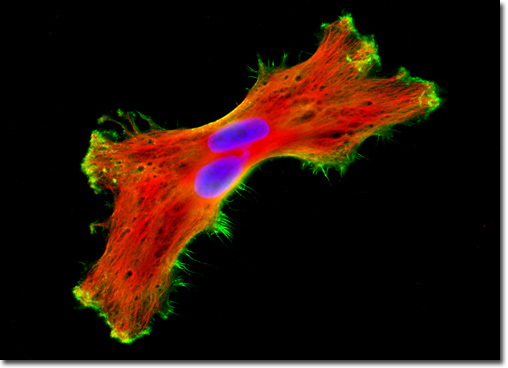
|
Texas Red is a hydrophilic sulfonylchloride derivative of the red-emitting fluorophore, sulforhodamine 101. The fluorescent probe was originally synthesized in the 1980s to provide a reagent for coupling to amino groups on proteins and other compounds. Texas Red emits at a longer wavelength (620 nanometers) than both tetramethylrhodamine and Lissamine rhodamine B, and exhibits only a minor amount of spectral overlap with fluorescein. Moreover, the fluorophore's fluorescence can be readily distinguished from that of the phycoerythrins. Thus, conjugates of the fluorophore are popular in the field of fluorescence microscopy as long-wavelength probes in multilabeling experiments. The single U-188 MG glioma cell presented in the digital image above was resident in a culture that was immunofluorescently labeled with anti-tubulin mouse monoclonal primary antibodies followed by goat anti-mouse Fab fragments conjugated to Texas Red. In addition, the specimen was stained with Alexa Fluor 488 conjugated to phalloidin and Hoechst 33342, targeting the cytoskeletal filamentous actin network and DNA in the cell nucleus, respectively. Images were recorded in grayscale with a QImaging Retiga Fast-EXi camera system coupled to an Olympus BX-51 microscope equipped with bandpass emission fluorescence filter optical blocks provided by Omega Optical. During the processing stage, individual image channels were pseudocolored with RGB values corresponding to each of the fluorophore emission spectral profiles. |
© 1995-2025 by Michael W. Davidson and The Florida State University. All Rights Reserved. No images, graphics, software, scripts, or applets may be reproduced or used in any manner without permission from the copyright holders. Use of this website means you agree to all of the Legal Terms and Conditions set forth by the owners.
This website is maintained by our
|