Fluorescence Digital Image Gallery
Mouse Embryo Teratocarcinoma Epithelial Cells (P19)
|
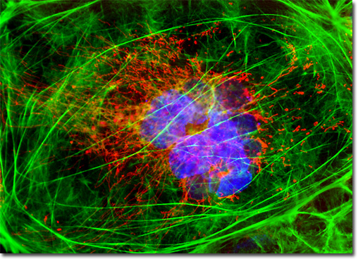
P19 cells, which exhibit epithelial morphology and adherent growth in culture, have been of significant interest to developmental biologists. Since the pluripotent murine embryonal carcinoma cells can differentiate in culture into many tissue types similar to those normally found in early embryos, they have they potential to help reveal important information about certain developmental proceedings. In some studies, for example, P19 cells have been used to analyze protein activities involved in neuronal differentiation. Other studies have concentrated more specifically on utilizing the P19 line as part of a larger endeavor to find ways that pluripotent cells may be applied in the medical field to develop treatments and cures for a variety of diseases and medical conditions, such as diabetes, Parkinson’s disease, and leukemia. The mouse embryo teratocarcinoma (P19) cell culture presented in the digital image above was labeled for mitochondria and the cytoskeletal filamentous actin network with MitoTracker Red CMXRos and Alexa Fluor 488 conjugated to phalloidin, respectively. In addition, the cells were counterstained for DNA with the ultraviolet-absorbing probe DAPI. Images were recorded in grayscale with a QImaging Retiga Fast-EXi camera system coupled to an Olympus BX-51 microscope equipped with bandpass emission fluorescence filter optical blocks provided by Omega Optical. During the processing stage, individual image channels were pseudocolored with RGB values corresponding to each of the fluorophore emission spectral profiles. |
© 1995-2025 by Michael W. Davidson and The Florida State University. All Rights Reserved. No images, graphics, software, scripts, or applets may be reproduced or used in any manner without permission from the copyright holders. Use of this website means you agree to all of the Legal Terms and Conditions set forth by the owners.
This website is maintained by our
|