Fluorescence Digital Image Gallery
Pig Kidney Epithelial Cells (LLC-PK1)
|
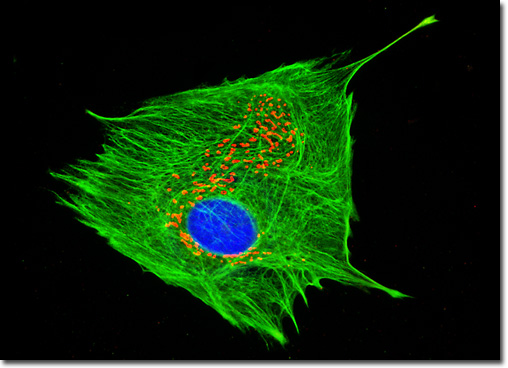
There has been a significant amount of scientific interest in porcine kidneys over the last few decades, chiefly because the organs have been widely viewed as the solution to the pronounced shortage of kidneys available for transplant purposes. Indeed, pig kidneys are considered better suited for xenotransplantation than those of many other animals due to their substantial anatomical and functional similarity to those of humans. In a pig, the left kidney is cranial to the right and the adrenal glands are located near the cranial poles of both of the organs. The renal blood supply is divided transversely between the cranial and caudal poles. Also, porcine kidneys are multirenculate and multipapillate, as are human kidneys. The organs are similar in size to those of human subjects as well when they are taken from the miniaturized breeds that are most prevalently used as laboratory animals. The log phase culture of pig kidney (LLC-PK1) epithelial cells presented in the digital image above was immunofluorescently labeled with anti-cytokeratin (pan) mouse monoclonal primary antibodies followed by goat anti-mouse Fab fragments conjugated to Oregon Green 488. Golgi bodies were simultaneously targeted with rabbit anti-giantin primary antibodies, followed by goat anti-rabbit secondaries conjugated to Texas Red. Nuclei were counterstained with Hoechst 33258. Images were recorded in grayscale with a QImaging Retiga Fast-EXi camera system coupled to an Olympus BX-51 microscope equipped with bandpass emission fluorescence filter optical blocks provided by Omega Optical. During the processing stage, individual image channels were pseudocolored with RGB values corresponding to each of the fluorophore emission spectral profiles. |
© 1995-2025 by Michael W. Davidson and The Florida State University. All Rights Reserved. No images, graphics, software, scripts, or applets may be reproduced or used in any manner without permission from the copyright holders. Use of this website means you agree to all of the Legal Terms and Conditions set forth by the owners.
This website is maintained by our
|