Microscopy Primer
Light and Color
Microscope Basics
Special Techniques
Digital Imaging
Confocal Microscopy
Live-Cell Imaging
Photomicrography
Microscopy Museum
Virtual Microscopy
Fluorescence
Web Resources
License Info
Image Use
Custom Photos
Partners
Site Info
Contact Us
Publications
Home

The Galleries:

Photo Gallery
Silicon Zoo
Pharmaceuticals
Chip Shots
Phytochemicals
DNA Gallery
Microscapes
Vitamins
Amino Acids
Birthstones
Religion Collection
Pesticides
BeerShots
Cocktail Collection
Screen Savers
Win Wallpaper
Mac Wallpaper
Movie Gallery

Simple Aquatic Microscope

The specimen, usually an aliquot of pond water, is placed in a watchglass that is held in the central circular beveled wooden stage. The model illustrated below was redrawn from photographs of the original microscope, which was photographed and described by Gerard Turner in his excellent catalog of microscopes from the Institute and Museum of the History of Science in Florence, Italy.

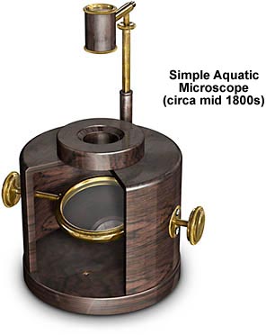
The body of the microscope consists of a wooden cylinder, painted black, that holds a substage mirror housed on an axle with knobs at the ends for position adjustment. A Coddington-style lens is mounted in a brass tube that is positioned with a short bracket attached to the microscope arm. The lens is made from a block of glass that has a v-shaped section cut around the center, and provides about 3x magnification. As discussed by Turner, the Coddington lens is named after Henry Coddington, a Cambridge University mathematics tutor, who published this design in 1829, although the actual inventor was David Brewster, who invented the lens in 1820.

BACK TO NINETEENTH CENTURY MICROSCOPES

Questions or comments? Send us an email.
© 1998-2022 by Michael W. Davidson and The Florida State University. All Rights Reserved. No images, graphics, scripts, or applets may be reproduced or used in any manner without permission from the copyright holders. Use of this website means you agree to all of the Legal Terms and Conditions set forth by the owners.
This website is maintained by our
Graphics & Web Programming Team
in collaboration with Optical Microscopy at the
National High Magnetic Field Laboratory.
Last modification: Friday, Nov 13, 2015 at 02:19 PM
Access Count Since January 8, 2000: 46635
For more information on microscope manufacturers,
use the buttons below to navigate to their websites: