
Peter Dollond Compound Monocular Eirometer
Originally designed to accurately measure thickness in threads of wool, the 3-inch long pocket-sized microscope possesses minimalist appeal. The model featured below was redrawn from photographs of the original microscope, which is part of the Billings microscope collection at Walter Reed Army Hospital in Washington DC.
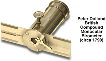
Comprised almost entirely of brass, the body screws into a Rochon micrometer and enables the microscopist to observe precise linear measurements of diameter and thickness. The magnifying power of the loupe-style compound eyepiece is assisted by the insertion of a field lens and an optional objective that screws into the micrometer plate. The circular stage positioned at the lower end of the body tube is rotated by a rack and pinion mechanism, and is equipped with double forceps that hold textile and hair specimens securely in place.
Designed to determine the thickness and size of wool fibers in industrial settings, this compact model was produced sometime around 1790. Sources of illumination most probably included sunlight, an oil lamp or indoor factory lighting. The microscope was manufactured by Peter Dollond in London, England -- a country that for centuries has been a leading producer of wool.
BACK TO EIGHTEENTH CENTURY MICROSCOPES