
Digital Image Capture and Processing
The Olympus MIC-D inverted digital microscope captures images with a complementary metal oxide semiconductor (CMOS) image sensor housed in the base and coupled to a host computer for acquisition, cataloging and processing of digital images.
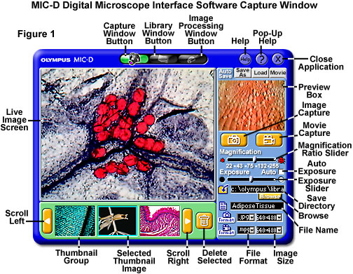
Interface software features include single image, full-motion video, and time-lapse sequence capture modes, in addition to a well-fortified cadre of image processing algorithms and an advanced image management system (see Figure 1). Digital images captured with the MIC-D microscope can be sorted, edited, corrected, and prepared for presentation, either electronically or through print media, with the accompanying software.
CMOS Image Sensors - The list of applications for CMOS image sensors has grown dramatically in the past several years. Since the late 1990s, CMOS sensors have accounted for increasing numbers of the imaging devices marketed in applications such as fax machines, scanners, security cameras, toys, games, PC cameras and low-end consumer cameras. The versatile sensors will also probably begin to appear in cell phones, bar code readers, optical mice, automobiles, and perhaps even domestic appliances in the coming years.
Basic Properties of Digital Images - Analog images captured by traditional photographic and microscopic techniques can be digitized and processed or viewed using personal computer equipment. This discussion addresses acquisition, sampling, and quantization of digital images as well as other important concepts, such as spatial resolution and dynamic range. In addition, color space models, image display technology, histograms, and storage requirements are also reviewed.
Fundamentals of Digital Image Processing - Digital image processing enables a reversible, virtually noise-free modification of an image in the form of a matrix of integers instead of the classical darkroom manipulations or filtration of time-dependent voltages necessary for analog images and video signals. Even though many image processing algorithms are extremely powerful, the average user often applies operations to digital images without concern for the underlying principles behind these manipulations. The result is often images that are severely degraded and compromised with respect to their potential, based on the power and versatility of the software utilized in the digital processing.
MIC-D Interface Software Capture Window - The Capture Window contains the live video feed from the MIC-D digital microscope CMOS image sensor. This software panel can be employed to capture single images, time-lapse sequence, or full-motion video sequences and save these image or audiovisual files to specific locations on the host computer's hard drive. In addition, images from previous microscopy sessions can be loaded into the Capture Window for comparison or editing purposes.
MIC-D Interface Software Image Processing Window - Single images captured by the MIC-D software, or other images saved in selected file formats, can be individually edited with a collection of image processing tools housed in the Image Processing Window. This window includes algorithms for performing operations such as resizing, cropping, rotation, color adjustments, sharpness, brightness, contrast, and gamma (color tone) on digital images.
MIC-D Interface Software Library Window - The MIC-D software Library Window can be utilized to view and select one or more images from a collection acquired during the current imaging session, or from a previously acquired group of images. This window is useful for comparing images (two or more), sorting through collections for individual images to rename or delete, viewing video sequence playbacks, and printing images.
Time-Lapse Cinemicrography - Time-lapse cinemicrography is a valuable tool for studying a wide variety of dynamic events, ranging from protracted particle motion and slowly recrystallizing chemicals to cellular movement and division. Many of these events occur over a period of many minutes or hours, a time scale that does not readily lend itself to being recorded with full-motion video. Instead, capturing sequential images at distinctly spaced time intervals followed by playback in real time can often enable the microscopist to observe relatively slow events on a much faster time scale.
Recommended Strategy for Processing Digital Images - Depending upon the illumination conditions, specimen integrity, and preparation methods, digital images captured in the optical microscope may require a considerable amount of rehabilitation to achieve a balance between scientific accuracy, cosmetic equilibrium, and aesthetic composition. When first acquired by a charge-coupled device (CCD) or complementary metal oxide semiconductor (CMOS) image sensor, digital images from the microscope often suffer from poor signal-to-noise, uneven illumination, focused or defocused dirt and debris, glare, color shifts, and a host of other ailments that degrade overall image quality.
Background Subtraction Toolkit - Because of the wide spectrum of illumination modes available with the MIC-D microscope, images can suffer from brightness variations that are manifested by gradients appearing in the background. These fluctuations often lead to contrast and brightness deficiencies in the specimen region and can seriously affect the quality of an otherwise acceptable digital image. This section discusses important details concerning the Background Subtraction Toolkit, which is designed to assist image processing applications by providing uniform backgrounds for specimens captured with the MIC-D digital microscope.
Background Subtraction Toolkit Download - The Molecular Expressions MIC-D digital microscope Background Subtraction Toolkit is a stand-alone Java application program designed for the Windows operating system, which can be utilized to produce uniform backgrounds for digital images captured with this unique inverted optical microscope. Use this link to visit the download area for additional information and to download the software to client computers.
Interactive Java Tutorials
MIC-D Digital Microscope Interface Software - The Olympus MIC-D digital microscope interface software contains three primary windows or menus that are utilized to capture, process, and catalog the images recovered from the microscope. This interactive tutorial explores the various functions of the interface software with a selection of single images, zoom sequences, and full-motion video produced by the actual software.
Background Subtraction - Application of a suitable background subtraction algorithm is a useful technique for correcting image defects that are associated with nonuniform brightness, often (but not always) attributed to uneven illumination in the microscope. This interactive tutorial explores a background subtraction image processing technique that relies on the creation of a background image from the original digital image.
Balancing Color in Digital Images - Color balancing belongs to an important class of digital image enhancement algorithms that are useful for correcting color casts in captured images. In most cases, unusual overall color casts (or uniform discolorations) typically result from color temperature effects in specimen illumination or are due to improper adjustment of the electronic detector (usually a digital CCD or CMOS camera system) used to capture the image.
Brightness and Contrast in Digital Images - The term contrast refers to the amount of color or grayscale differentiation that exists between various image features in both analog and digital images. Images having a higher contrast level generally display a greater degree of color or grayscale variation than those of lower contrast. Image brightness (or luminous brightness) is a measure of intensity after the image has been acquired with a digital camera or digitized by an analog-to-digital converter. This interactive tutorial explores the wide range of adjustment that is possible in digital image brightness and contrast manipulation, and how these variations affect the final appearance of the image.
Convolution Kernel Mask Operation - A powerful array of image-processing methods utilize multipixel operations, in which each output pixel is altered by contributions from a number of adjoining input pixels. These types of operations are referred to as convolution or spatial convolution. This interactive tutorial explores how a convolution operation is performed on a digital image.
Gamma Correction - The perceived brightness of a digital image captured with an optical microscope is dependent not only upon the conditions of specimen illumination, but also on the sensitivity and linearity of the detector upon which the image was gathered. In addition, the characteristics of the display device (television, computer monitor, flat-screen display) where the digital image is viewed also affect the intensity distribution and interrelationship of contrast between light and dark regions in the specimen. The effects are characterized by a variable known as gamma, which is explored in this interactive tutorial.
Gray-Level Resolution - The Olympus MIC-D digital microscope is equipped with a CMOS image sensor that captures images having 640 x 480 pixel dimensions in red, green, and blue (RGB) true color mode with 8-bit resolution per channel, resulting in a total of 256 tonal levels for each of the three colors. When the color images are converted to grayscale by an image-editing software program, they produce corresponding black and white (monochrome) images that have a single 8-bit channel with 256 gray levels.
JPEG Image Compression - The JPEG lossy image compression standard is currently in worldwide use, and is becoming a critical element in the storage of digital images captured with the optical microscope. This interactive tutorial explores compression of digital images with the JPEG algorithm, and how the lossy storage mechanism affects file size and the final image appearance.
Adjustment of Digital Image Sharpness - The sharpness of a digital image refers to the degree of clarity in both coarse and fine specimen detail. A lack of sharpness in digital images captured with the microscope often results from poor focus adjustment, vibration, or the specimen not being flat with respect to the imaging plane. This common artifact can also result from a variety of optical aberrations such as spherical aberration, astigmatism, coma, geometrical distortion, and field curvature.
Digital Image Sampling Frequency - In order to match the optical and electronic resolution of a microscope and the accompanying camera system, a digital image should have a sufficient number of samples per horizontal line so that the display faithfully represents the original signal presented to the digitizing device. This interactive tutorial explores how variations in specimen sampling frequency affect the resolution of the final image.
Spatial Resolution - Spatial resolution is a term that refers to the number of pixels utilized in construction of a digital image. Images having higher spatial resolution are composed with a greater number of pixels than those of lower spatial resolution. This interactive tutorial explores variations in digital image spatial resolution, and how these values affect the final appearance of the image.
Cropping, Zooming, and Repositioning Digital Images - The microscopist must often arrange digital images on the output display device for purposes of visually comparing image details, or for illustrating differences between two or more images. Such display operations are often facilitated through software packages that enable interactive panning, scrolling, flipping, scaling, and zooming of digital images. Zooming to enlarge image details is useful for the visualization of small structures present in a digital image, and scaling is often necessary to format a digital image to fit within the boundaries of a display medium, as in the case of displaying a collection of thumbnail images.
Unsharp Mask Filtering - Enhancing the overall sharpness of a digital image often has the effect of revealing fine details that cannot be clearly discerned in the original. The unsharp mask filter algorithm is an extremely versatile sharpening tool that improves the definition of fine detail by removing low-frequency spatial information from the original image. This interactive tutorial explores how the unsharp mask filter algorithm is utilized to sharpen details in a variety of digital images captured with the Olympus MIC-D digital microscope.
White and Black Balance - The color of digital images captured with an optical microscope is dependent not only upon the spectrum of visible light wavelengths transmitted through or reflected by the specimen, but also on the spectral content of the illuminator. In color digital camera systems that employ either charge-coupled device (CCD) or complementary metal oxide semiconductor (CMOS) image sensors, white and/or black balance (baseline) adjustment is often necessary in order to produce acceptable color quality in digital images.
Contributing Authors
Kenneth R. Spring - Scientific Consultant, Lusby, Maryland, 20657.
John C. Russ - Materials Science and Engineering Department, North Carolina State University, Raleigh, North Carolina, 27695.
Thomas J. Fellers and Michael W. Davidson - National High Magnetic Field Laboratory, 1800 East Paul Dirac Dr., The Florida State University, Tallahassee, Florida, 32310.
BACK TO THE OLYMPUS MIC-D DIGITAL MICROSCOPE
Questions or comments? Send us an email.
© 1995-2025 by Michael W. Davidson and The Florida State University. All Rights Reserved. No images, graphics, software, scripts, or applets may be reproduced or used in any manner without permission from the copyright holders. Use of this website means you agree to all of the Legal Terms and Conditions set forth by the owners.
This website is maintained by our
Graphics & Web Programming Team
in collaboration with Optical Microscopy at the
National High Magnetic Field Laboratory.
Last Modification Friday, Nov 13, 2015 at 02:19 PM
Access Count Since September 17, 2002: 44731
Visit the website of our partner in introductory microscopy education:
|
|