
The Olympus MIC-D Digital Microscope
Olympus has thrown the doors open to a new era in optical microscopy education with the introduction of the MIC-D inverted digital microscope. Designed specifically for a wide spectrum of applications ranging from basic classroom instruction to more advanced laboratory analysis, this versatile microscope features a palette of contrast enhancing techniques that rival many research-level instruments.
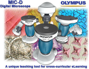
Anatomy of the MIC-D Digital Microscope - The Olympus MIC-D digital microscope represents a unique design that incorporates a CMOS electronic digital imaging sensor as a substitute for the traditional eyepieces or oculars found in a majority of microscopes. Coupled to an inverted illumination system that broadcasts light from above the specimen (similar to a tissue culture microscope), this digital microscope also features a translatable lamphouse and condenser unit that can be rotated over a 135-degree angle. Such versatility allows the operator to project light onto the specimen from an almost limitless combination of oblique or reflected illumination angles.
Digital Image Capture and Processing - The Olympus MIC-D inverted digital microscope captures images with a complementary metal oxide semiconductor (CMOS) image sensor housed in the base and coupled to a host computer for acquisition, cataloging and processing of digital images. Interface software features include single image, full-motion video, and time-lapse sequence capture modes, in addition to a well-fortified cadre of image processing algorithms and an advanced image management system. Digital images captured with the MIC-D microscope can be sorted, edited, corrected, and prepared for presentation, either electronically or through print media, with the accompanying software.
Software Interface Online Demonstration - The Olympus MIC-D digital microscope interface software contains three primary windows or menus that are utilized to capture, process, and catalog the images recovered from the microscope. This interactive tutorial explores the various functions of the interface software with a selection of single images, zoom sequences, and full-motion video produced by the actual software.
MIC-D Digital Microscope Quick Start Guide (PDF Format; 5.2 Mb) - Download the official Olympus MIC-D Quick Start Guide, which provides a brief explanation of the microscope hardware and software, as well as hints on specimen choice, illumination techniques, and image processing. In the laboratory or in the field, the Olympus MIC-D portable digital microscope captures still and moving images instantly. The guide will introduce beginners to the microscope's innovative design and spectacular imaging capabilities, enabling them to collect images on the microscope with a minimum of effort.
Digital Image Galleries - The Olympus MIC-D digital microscope image galleries contain a wide spectrum of images representing all of the illumination techniques available with this unique instrument. Specimens include stained thin sections, whole mounts, thick sections, living pond creatures, insects, recrystallized chemicals, and integrated circuits. Imaging of specimens contained in the gallery was assisted by the various contrast-enhancing technology featured by the MIC-D, including brightfield, darkfield, oblique, polarized light and reflected illumination, and the galleries are arranged according to contrast technique.
Digital Video Galleries
Collections of time-lapse sequences featuring common chemical crystals undergoing melting transitions and pond life captured with the MIC-D in full-motion video are presented in this gallery. Video sequences are presented either as interactive Java tutorials or in Windows Media Player, and available in several playback formats to match a variety of Internet connection speeds.
Chemical Crystals - Chemical compounds can exist in three basic phases: gaseous, liquid, or solid. Gases consist of weakly bonded atoms and expand to fill any available space. Solids are characterized by strong atomic bonding and have a rigid shape. Most are crystalline, having a three-dimensional periodic atomic arrangement. Some, such as glass, lack this periodic arrangement and are noncrystalline, or amorphous. Liquids have characteristics that fall in between gases and solids. This cinemicrographic collection shows time-lapse movies of various chemical compounds as they change physical states.
Pond Life - Freshwater ponds provide a home for a wide variety of aquatic and semi-aquatic plants, insects, and animals. The vast majority of pond inhabitants, however, are invisible until viewed under the microscope. Beneath the placid surface of any pond is a microscopic metropolis bustling with activity as tiny bizarre organisms pursue their lives; locomoting, eating, trying not to be eaten, excreting, and reproducing. In this collection of digital movies, observe the activities of microscopic organisms taken from a typical North Florida pond.
MIC-D Digital Microscope Screen Savers
MIC-D Digital Microscope Screen Savers - Visitors who enjoy the galleries are invited to download and install screen savers containing images from the galleries. Our software programmers have worked in collaboration with microscopists to develop a series of screen savers using digital images captured with the MIC-D microscope operating in a variety of contrast-enhancing modes. These screen savers have been developed for computers utilizing the Windows (95, 98, NT, 2000, and XP) operating system and are available as free downloads to our visitors. We hope you enjoy these screen savers, which are provided at no charge by Olympus America, Inc. and The Florida State University.
Contributing Authors
Kenneth R. Spring - Scientific Consultant, Lusby, Maryland, 20657.
John C. Russ - Materials Science and Engineering Department, North Carolina State University, Raleigh, North Carolina, 27695.
Matthew Parry-Hill, Christopher A. Burdett, Thomas J. Fellers, Cynthia D. Kelly, Omar Alvarado, John D. Homan and Michael W. Davidson - National High Magnetic Field Laboratory, 1800 East Paul Dirac Dr., The Florida State University, Tallahassee, Florida, 32310.
Questions or comments? Send us an email.
© 1995-2025 by Michael W. Davidson and The Florida State University. All Rights Reserved. No images, graphics, software, scripts, or applets may be reproduced or used in any manner without permission from the copyright holders. Use of this website means you agree to all of the Legal Terms and Conditions set forth by the owners.
This website is maintained by our
Graphics & Web Programming Team
in collaboration with Optical Microscopy at the
National High Magnetic Field Laboratory.
Last Modification Friday, Nov 13, 2015 at 02:18 PM
Access Count Since September 17, 2002: 327458
Visit the website of our partner in introductory microscopy education:
|
|