Microscopy Primer
Light and Color
Microscope Basics
Special Techniques
Digital Imaging
Confocal Microscopy
Live-Cell Imaging
Photomicrography
Microscopy Museum
Virtual Microscopy
Fluorescence
Web Resources
License Info
Image Use
Custom Photos
Partners
Site Info
Contact Us
Publications
Home

The Galleries:

Photo Gallery
Silicon Zoo
Pharmaceuticals
Chip Shots
Phytochemicals
DNA Gallery
Microscapes
Vitamins
Amino Acids
Birthstones
Religion Collection
Pesticides
BeerShots
Cocktail Collection
Screen Savers
Win Wallpaper
Mac Wallpaper
Movie Gallery

Polarized Light Microscopy Digital Image Gallery

Micropegmatite Granite

Graphic granite is a type of granite that lacks mica and is chiefly composed of quartz and feldspar. The crystals in the rock are arranged in such a way that in transverse sections they appear similar to oriental characters or runes, a fact that is responsible for the rock’s alternate moniker, runite.

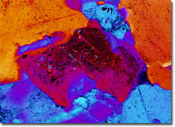
When the unusual crystal structure of graphic granite occurs on a microscopic scale, the rock is usually referred to as micropegmatite, though it is also occasionally known by the name of microgranite. This structure is specifically comprised of patches of quartz crystals in parallel orientation, which usually appear triangular in cross section, and quartz-feldspar interfaces that are planar. The remarkable angularity and parallelism of the crystals of quartz are generally believed to be a reaction to the conditions present during their growth.

Micropegmatite is the dominant material that comprises granophyres, fine-grained granite porphyries that consist of irregular intergrowths of quartz and feldspar imbedded in a groundmass. However, the rock may also often be found among common granites and the fine-grained igneous rocks called diabases. Within the latter, micropegmatite is often interstitial, appearing as veins, seams, or aggregates.


BACK TO THE ROCKS AND MINERALS GALLERY

BACK TO THE POLARIZED LIGHT GALLERY

Questions or comments? Send us an email.
© 1998-2022 by Michael W. Davidson and The Florida State University. All Rights Reserved. No images, graphics, scripts, or applets may be reproduced or used in any manner without permission from the copyright holders. Use of this website means you agree to all of the Legal Terms and Conditions set forth by the owners.
This website is maintained by our
Graphics & Web Programming Team
in collaboration with Optical Microscopy at the
National High Magnetic Field Laboratory.
Last modification: Friday, Nov 13, 2015 at 02:19 PM
Access Count Since November 20, 2003: 14234
For more information on microscope manufacturers,
use the buttons below to navigate to their websites: