Microscopy Primer
Light and Color
Microscope Basics
Special Techniques
Digital Imaging
Confocal Microscopy
Live-Cell Imaging
Photomicrography
Microscopy Museum
Virtual Microscopy
Fluorescence
Web Resources
License Info
Image Use
Custom Photos
Partners
Site Info
Contact Us
Publications
Home

The Galleries:

Photo Gallery
Silicon Zoo
Pharmaceuticals
Chip Shots
Phytochemicals
DNA Gallery
Microscapes
Vitamins
Amino Acids
Birthstones
Religion Collection
Pesticides
BeerShots
Cocktail Collection
Screen Savers
Win Wallpaper
Mac Wallpaper
Movie Gallery

Compound Monocular Tripod Microscope

The body of this microscope is 24 inches when fully extended and 16 inches when closed. John Mayall obtained this microscope in France for Colonel Billings in 1887, where it was reported to have been in the possession of one family since early in the 1700's. The model featured below was redrawn from photographs of the original microscope, which is part of the Billings microscope collection at Walter Reed Army Hospital in Washington DC.

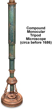
Covered with green vellum and gold stampings, the body of this beautiful microscope is fashioned from pasteboard and contains three draw tubes. The wooden (probably pear) circular base has three legs that support a turned wood cone socket that secures the body tube. Focusing is achieved by sliding the draw tubes in and out. At the top of the lowest tube is a 7/8-inch diameter field lens, and the draw tube, which carries the eye lens, slides over the body tube, thus varying the distance between the two lenses. The objective is mounted in a wooden fixture that screws into the nosepiece.

BACK TO SIXTEENTH-SEVENTEENTH CENTURY MICROSCOPES

Questions or comments? Send us an email.
© 1998-2022 by Michael W. Davidson and The Florida State University. All Rights Reserved. No images, graphics, scripts, or applets may be reproduced or used in any manner without permission from the copyright holders. Use of this website means you agree to all of the Legal Terms and Conditions set forth by the owners.
This website is maintained by our
Graphics & Web Programming Team
in collaboration with Optical Microscopy at the
National High Magnetic Field Laboratory.
Last modification: Friday, Nov 13, 2015 at 02:19 PM
Access Count Since December 8, 2000: 44786
For more information on microscope manufacturers,
use the buttons below to navigate to their websites: