Microscopy Primer
Light and Color
Microscope Basics
Special Techniques
Digital Imaging
Confocal Microscopy
Live-Cell Imaging
Photomicrography
Microscopy Museum
Virtual Microscopy
Fluorescence
Web Resources
License Info
Image Use
Custom Photos
Partners
Site Info
Contact Us
Publications
Home

The Galleries:

Photo Gallery
Silicon Zoo
Pharmaceuticals
Chip Shots
Phytochemicals
DNA Gallery
Microscapes
Vitamins
Amino Acids
Birthstones
Religion Collection
Pesticides
BeerShots
Cocktail Collection
Screen Savers
Win Wallpaper
Mac Wallpaper
Movie Gallery

Phase Contrast Image Gallery

Black Grape Rot

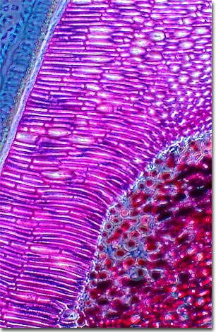
Black rot, caused by the fungus Guignardia bidwellii is one of the most serious diseases of cultivated grapes in the eastern United States, especially in warm, humid areas. As evidenced by this micrograph, combining phase contrast microscopy with classical histological staining techniques often yields enhancement of cellular features.

Crop losses can be devastating, ranging from 5-80% depending on the weather, the variety of grape being grown, and the amount of disease in the vineyard. The fungus can infect all green parts of the vine but the most damaging effect is on the fruit, which shrivel up into dark-colored mummies.

This fungus reproduces with two types of spores, ascospores and conidia (pycniospores). Ascopores are produced in the grape mummy and forcibly discharged into the air, often traveling considerable distances. Conidia are vegetatively reproduced spores that the fungus uses to propagate, and are spread through rain or irrigation water splashing on the plants.

VIEW THIS SPECIMEN UNDER FLUORESCENCE ILLUMINATION

BACK TO THE PHASE CONTRAST GALLERY

Questions or comments? Send us an email.
© 1998-2022 by Michael W. Davidson and The Florida State University. All Rights Reserved. No images, graphics, scripts, or applets may be reproduced or used in any manner without permission from the copyright holders. Use of this website means you agree to all of the Legal Terms and Conditions set forth by the owners.
This website is maintained by our
Graphics & Web Programming Team
in collaboration with Optical Microscopy at the
National High Magnetic Field Laboratory.
Last modification: Friday, Nov 13, 2015 at 02:19 PM
Access Count Since March 15, 2000: 27669
For more information on microscope manufacturers,
use the buttons below to navigate to their websites: