Phase Contrast Image Gallery
Agatized Dinosaur Bone
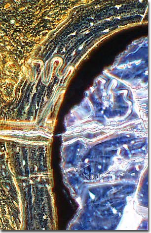
Agate crystalline formation within the Haversian canals is quite evident in this photomicrograph, which was taken during examination of a thin section prepared from fossilized dinosaur bone fragments.

Fossilized dinosaur bones come in a variety of forms depending on how they have been petrified. Most dinosaur bones have been petrified with calcium, which yields a stony appearance and texture. Agatized bones are petrified with silica, or quartz crystals, giving them a colorful, glassy appearance.
Fossilization occurs when hard parts from a dead plant or animal are buried quickly in sediments and exposed to mineralized solutions over long periods of time. Minerals dissolved in ground waters infiltrate the organic tissues, filling the spaces within and between cells, gradually embedding and preserving the tissue structure. On rare occasions, even soft body parts, eggs, and feces have been fossilized.
For bones to become agatized, a process called permineralization occurs in which groundwater, rich in silicon dioxide (silica), infiltrates the bone. The mineral rich solution fills the spaces of the bone with silica crystals, eventually forming patterns of colorful quartz.
Agatized bones are only found in areas of the world that were rich with silica minerals at the time the bones were fossilizing. In the United States, the largest deposits of agatized dinosaur bones are concentrated in Colorado and Utah.
BACK TO THE PHASE CONTRAST GALLERY
