
Hoffman Modulation Contrast Image Gallery
Down Feathers
Feathers are excellent candidates for photography throught the microscope by standard brightfield techniques, darkfield, polarized light, and/or differential interference contrast. The image below is a photomicrograph of down feathers illuminated with a combination of polarized light and Hoffman modulation contrast.
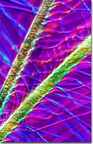
Down feathers, like all other feathers, are specialized epidermal growths, formed by papillae, that are composed of a fibrous protein named keratin (about 90%), lipids, and pigments that give them their beautiful colors. Keratin is an ideal material for feathers because it is lightweight and flexible, yet strong enough to form a structure that can withstand the rigors of flight.
Feathers can be divided into several categories. Contour feathers are found on the wings and backs of birds, while down feathers are usually placed underneath the contour feathers to provide insulation for the bird. A primary feature present in all feathers is the large stem (termed the "rachis" or "quill") centrally located that provides a "backbone" around which the feather is constructed. Extending from both sides of the quill is a linear cluster of barbs containing a majority of the feather's pigment. Together, the barbs and the quill constitute the body or vane of the feather. Branching at a 40-degree angle on each barb is a network of barbules with interlocking hooklets that provide both stiffness and flexibility to the feather. These hooklets allow a split vane to be resealed making it whole again.