
Differential Interference Contrast Image Gallery
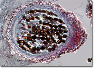
Fungus (Sordaria fimicola) Fruiting Bodies
Sordaria fimicola is an ascomycete fungus that normally grows on decaying organic material. It may also be frequently found in introductory laboratory settings where it is manipulated and examined for educational purposes.
Ascomycetes are known as sac fungi because of the characteristic shape of their asci, which each contain four to eight ascospores in the sexual stage. The specific attributes of the asci and the method of release of the ascospores is what primarily determines which subgroup ascomycete species are placed in. Other differences do exist between the fungi, however, which may follow very different paths of existence. Some ascomycetes are pathogens that cause disease in plants or animals, while others are edible or harmlessly live on dead organic matter. Perhaps the ascomycete most important to man is Saccharomyces cerevisiae, the common yeast that is used around the world to leaven bread and ferment the grain that produces beer.
The popularity of using S. fimicola as an instructional tool is largely due to its simplicity to work with. The fungus grows well in culture and can produce mature perithecia, or fruiting bodies, in about a week. Students can then easily observe the meiotic division of the diploid perithecia, which produce ordered linear tetrads encased in the ascus sac. Subsequently, these tetrads are converted to octads by mitosis of the haploid ascospores, providing students with the opportunity to learn about another important biological process.