
Differential Interference Contrast Image Gallery
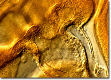
Chicken Embryo
The chicken has been domesticated for approximately 8,000 years and, over that time, has been exploited for a wide variety of purposes. Egg and meat production, cockfighting, feather yield, and scientific study are a few of the most common reasons the chicken has been of such significant interest to man for so many years.
In the laboratory, chicken embryos and cultured cells are of great use to scientists. Most chickens have genes that are similar to those found in humans and, therefore, chicken cells may be utilized to achieve a better understanding of human biology and mammalian development in general. Chicken embryos are normally better suited to controlled studies than other laboratory animals, such as mice and rats, because they develop inside an egg rather than in the body of the mother. In fact, the relatively recent increase in the scientific use of chicken embryos may help reduce animal testing by replacing exposure trials on mice and rats.
The full development of the chicken embryo is a multi-step process that takes weeks to complete. After the mating of two mature adult chickens, the female’s eggs may be fertilized within 24 hours. Yolks begin to develop in the ovary and then are passed into the oviduct once they reach a diameter of approximately 4 centimeters. There two shell membranes, structural fibers, and the thick egg white, or albumin, are added. As the egg travels further down the oviduct it continually rotates through the spiraling tube causing the structural fibers to twist into rope-like structures known as chalazae that anchor the yolk to the thick egg white. In the uterus, just prior to the laying of the egg, a thin layer of egg white is added and a calcite eggshell is deposited around the egg. Once a fertilized egg is laid, the chicken embryo grows and develops for approximately 21 days before it is ready to hatch.