
The New Universal Single Microscope
In 1746 George Adams announced the New Universal Single microscope, along with the New Universal Double microscope, in his Micrographia Illustrata.
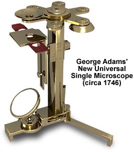
Adams created the Universal Single microscope with the intention of producing a microscope with both portability and a wide range of applications. The basic microscope consisted of six bi-convex lenses mounted on a rotating wheel that was a precursor to the modern revolving nosepiece. The lenses could be all rotated in sequence under the stationary eye shield to achieve successively higher degrees of magnification. One of the lenses was mounted with a Lieberkuhn reflector, which Adams termed "a reflecting speculum of silver or other metal, highly polished" that was very useful for aiding illumination of opaque specimens.
The Universal Single used a convenient focusing technique that consisted of a knurled adjustment wheel located at the bottom center of the pillar (visible in the illustration above). This wheel was attached to a screw that traversed the interior of the pillar and was connected to the stage arm. As the focusing wheel was turned, the resulting turning of the interior screw either pulled or pushed the stage arm bringing the sample into focus. As a useful indicator, graduation lines were engraved onto the exterior of the pillar with corresponding numbers to show the various focal points of the six different lenses.
BACK TO EIGHTEENTH CENTURY MICROSCOPES