
English Tripod Microscope
Some of the earliest known British microscopes consist of a concentric set of cylindrical tubes capped on one end with an eye lens cup and tapering on the other into a threaded nosepiece containing an objective lens, the entire assembly being supported by a set of tripod legs.
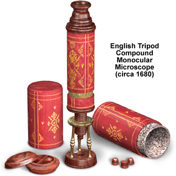
The microscope illustrated above was redrawn from a photograph of a late seventeenth century replica from the collection of Dr. James B. McCormick. It is uncertain who actually designed and originally built this microscope, but the most likely candidate is British instrument maker and optician John Yarwell. The outer body tube is made of pasteboard covered with red morocco, while the inner tube is pasteboard covered with green vellum. Both tubes are decorated with ornate gold tooling, much like book bindings of the period. Three lenses are used to image specimens: A single lens in the eyepiece, a bi-convex field lens located at the top of the inner body tube, and an interchangeable objective lens in a wooden mount that is screwed into the end of the nosepiece. The circular wooden box to the left of the microscope contains storage room for four wood-mounted objectives and the entire assembly screws onto the top of the eyepiece when the microscope is packed away.
Focus is achieved by screwing the two-inch long threaded nosepiece in and out of the tripod collar. The microscope stand is made from a very dark hardwood that has been fashioned into a circular base upon which a brass tripod is mounted. Samples are placed over the hole in the base and the microscope is then held up to a light source for transmitted microscopy. Opaque samples are placed on top of the ivory insert (shown on the base), which is inserted into the base hole.
BACK TO SIXTEENTH-SEVENTEENTH CENTURY MICROSCOPES