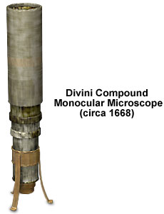
Divini Compound Monocular Microscope
Eustachio Divini, the great Italian optics pioneer and telescope maker, designed the compound monocular microscope illustrated below in about 1668. A reproduction model was commissioned in 1888 by British instrument maker and microscope collector John Mayall, and resides in the Billings Microscope Collection at the National Museum of Health and Medicine at Walter Reed Army Hospital in Washington, D. C.

The original microscope is housed in the Museo Astronomico e Copernicano in Rome, Italy, and is also described in McCormick and Turner's book, The Atlas Catalogue of Replica Rara Ltd. Antique Microscopes. Divini also initially published a description of the microscope in the Giornale dei Letterati. Two plano-convex lenses, which are arranged with their curved surfaces in contact to produce a flatter field, comprise the compound eyepiece. The range of magnification is described as 41 to 143 diameters, and is adjusted with four telescoping draw tubes. A description of the objective is lacking, but it is probably a bi-convex lens similar to the 7/16-inch diameter objective featured in Divini's 1670 wood and brass microscope. At the upper end of the instrument, a diaphragm is included for controlling illumination and specimen resolution.
Cardboard that is covered with gray paper was utilized to wrap the microscope body (as illustrated). However, age and oxidation have taken their toll on both the Divini original in Rome and the reproduction in Washington, D.C., and the paper has turned brown, according to Blumberg and his co-authors in the description of the Billings collection. The tripod base with bent feet, and the cylindrical socket that holds the microscope body, are made of tin. Each draw tube is inscribed with its magnification, and features an external collar at the lower end that acts as a stop. The bottom or smallest draw tube carries the objective lens in a tin mounting frame. Fully extended, the Divini compound microscope measures 16-1/2-inches tall and the diameter of the upper tube is 1-1/2 inches.
BACK TO SIXTEENTH-SEVENTEENTH CENTURY MICROSCOPES
