
Top Hat Filter
The top hat filter is based on neighborhood ranking, but it uses the ranked value from two different size regions. The brightest value in a circular interior region is compared to the brightest value in a surrounding annular region. If the brightness difference exceeds a threshold level, it is kept (otherwise it is erased). This interactive tutorial illustrates the application of a top hat filter to isolate details in an image.
The tutorial initializes with a randomly selected specimen appearing in the Specimen Image window. The Choose A Specimen pull-down menu provides a selection of specimen images, in addition to the initial randomly chosen one. The Filtering Mode buttons select whether to keep pixels that are Bright or Dark compared to their surroundings. The Neighborhood Radius slider adjusts the size of the interior region used in the top hat comparison (the outer or annular region is one or two pixels wide). The Threshold slider controls the difference between the brightest or darkest pixel value in the interior and surrounding regions that must be exceeded for the pixel to be retained, to produce the resulting Filtered Image shown on the right.
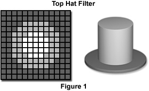
If the interior and annular regions are drawn as shown in the diagram in Figure 1, the reason for the filter name becomes apparent. The interior region is the crown and the threshold is its height, while the surrounding annulus is the brim of the hat. This operation is particularly well suited for finding the spikes in Fourier transform power spectra.
Contributing Authors
John C. Russ - Materials Science and Engineering Dept., North Carolina State University, Raleigh, North Carolina, 27695.
Matthew Parry-Hill, and Michael W. Davidson - National High Magnetic Field Laboratory, 1800 East Paul Dirac Dr., The Florida State University, Tallahassee, Florida, 32310.
BACK TO INTRODUCTION TO DIGITAL IMAGE PROCESSING AND ANALYSIS
BACK TO MICROSCOPY PRIMER HOME