
Laplacian Sharpening
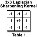
Sharpening of images to increase local contrast is almost universally applied by publishers to counter the visual blurring effect of halftoning images in the printing process. This is usually done by a convolution using a kernel of weights, often generated by a Gaussian smoothing function. But in this application, some of those weights will have negative values. For instance, the Laplacian sharpening filter in Table 1 combines each pixel with its eight adjacent neighbors. This interactive tutorial illustrates the process of Laplacian sharpening using the 3x3 kernel illustrated below.
The tutorial initializes with a randomly selected specimen appearing in the Specimen Image window. The Choose A Specimen pull-down menu provides a selection of specimen images, in addition to the initial randomly chosen one. Adjacent to the Specimen Image window is the Laplacian Image window showing the result of applying a Laplacian filter to the specimen. In the tutorial, the sharpening is applied only to the pixel brightness values, retaining the original color information.
Contributing Authors
John C. Russ - Materials Science and Engineering Dept., North Carolina State University, Raleigh, North Carolina, 27695.
Matthew Parry-Hill, and Michael W. Davidson - National High Magnetic Field Laboratory, 1800 East Paul Dirac Dr., The Florida State University, Tallahassee, Florida, 32310.
BACK TO INTRODUCTION TO DIGITAL IMAGE PROCESSING AND ANALYSIS
BACK TO MICROSCOPY PRIMER HOME