
Directional Derivatives
The boundaries of features and structural details in an image are usually defined by changes in brightness or color. Locating these edges so that they can be accurately measured has been an active area of development in image processing. In addition, there is considerable evidence that the steps and edges are key components of a scene selected by human visual processes, and that either extracting them from an image (forming a “sketch” of the scene) or sharpening their appearance assists viewers.
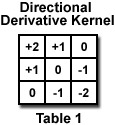
The Laplacian sharpening or unsharp mask filters shown above increase the contrast at steps and edges, but are also sensitive to noise. These operations are mathematically a second derivative of brightness. A first derivative of brightness can be calculated with a kernel such as the one in Table 1 that calculates the difference between neighbors at the upper left and lower right. Because it can produce either positive or negative results, an offset value of 128 (medium gray) is added to the result for display. Eight similar kernels can be formed by rotating the values in 45 degree steps to calculate the derivative in various directions. This interactive tutorial illustrates the application of a directional derivative filter to enhance the visibility of details in an image.
The tutorial initializes with a randomly selected specimen appearing in the Specimen Image window. The Choose A Specimen pull-down menu provides a selection of specimen images, in addition to the initial randomly chosen one. Adjacent to the Specimen Image window is the Output Image window showing the result of applying a directional derivative filter to the specimen. The Edge Direction slider rotates the directional derivative filter shown, to produce the resulting Filtered Image shown on the right.
Contributing Authors
John C. Russ - Materials Science and Engineering Dept., North Carolina State University, Raleigh, North Carolina, 27695.
Matthew Parry-Hill, and Michael W. Davidson - National High Magnetic Field Laboratory, 1800 East Paul Dirac Dr., The Florida State University, Tallahassee, Florida, 32310.
BACK TO INTRODUCTION TO DIGITAL IMAGE PROCESSING AND ANALYSIS
BACK TO MICROSCOPY PRIMER HOME