Recommended Books on Optical Microscopy
Several hundred books dealing with various aspects of optical microscopy and related fields are currently available from the booksellers. This section lists the Molecular Expressions website team top 10 recommended books on microscopy, digital imaging, fluorescence, video, and microtechnique.
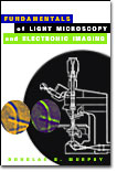
Fundamentals of Light Microscopy and Digital Imaging - Douglas B. Murphy. Professor Murphy constructs a solid foundation on the basic concepts of geometrical optics, light, and color, and then provides excellent introductory reviews of important topics in light microscopy. The book is very well written and complex phenomena are clearly explained without the unnecessary math that often confuses students. Illustrations are numerous and help support the text very nicely, as do the suggested laboratory exercises that accompany each chapter. Discussions of digital cameras and image processing are timely and provide the essential concepts necessary to tackle more advanced treatises. In the opinion of the Molecular Expressions microscopy website team, this book is by far the best entry-level textbook in the field. For students and researchers who can only afford a single volume on microscopy, Professor Murphy's book is definitely the best choice.
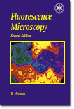
Fluorescence Microscopy, 2nd Edition - Brian Herman. Fluorescence Microscopy is the revised, updated and expanded second edition of a best-selling introductory text by professor Herman on the use of fluorescence microscopy in cell biology and related arenas. The book covers the fundamental principles of fluorescence, configuration and operation of the microscope, as well as the application of these concepts to practical fluorescence microscopy. In addition, a wide spectrum of advanced techniques, including FISH, TIRFM, FRET, FLIM, fluorescence/DIC, image cytometry, recovery after photobleaching, and polarization anisotropy are also discussed. Introductory principles of quantitative fluorescence microscopy are presented in a clear and concise manner and a discussion of photomicrography and digital imaging techniques help introduce the reader to these necessary methods for image capture. Troubleshooting guidance is provided, along with many practical hints, to guide the user through commonly encountered problems. This volume is an ideal introduction to the topic of fluorescence microscopy for beginning students and researchers.
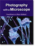
Photography with a Microscope - Fred Rost and Ron Oldfield. Starting with the basics of photomicrography, Rost and Oldfield present a detailed description of the microscope and review a host of specialized optical techniques. Included is introductory information on darkfield, polarized light, stereomicroscopy, phase contrast, differential interference contrast (DIC), fluorescence, infrared, and ultraviolet microscopy procedures. Later sections review film camera systems, including the complex topic of exposure determination and measurement, and advanced topics in photomicrography. Digital and video techniques are reviewed and, although much of the photographic methodology is rapidly being supplanted by digital techniques, the volume offers a handy reference guide for those who currently employ traditional film cameras. Several appendices discuss care and maintenance of microscope and camera equipment, and offer extensive troubleshooting tips to guide the reader when things go wrong. The reference section is also a valuable catalog of books and review articles that deal with the marriage of cameras to microscopes.
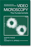
Video Microscopy: The Fundamentals - Kenneth R. Spring and Shinya Inoué. Although many of the video techniques and tube cameras discussed by Drs. Spring and Inoué have largely been replaced by computers and digital camera systems, the book is still one of the best reference guides to basic concepts in optical microscopy. Chapter 2 (Microscope Image Formation) presents a solid review of the complex topics involved in forming images with diffraction-limited optical systems and relates these concepts to the microscope. In addition, specialized topics are discussed, as well as practical aspects of everyday microscopy, including Köhler illumination, microscope alignment, light sources, image brightness, and lens choice. An excellent review of the physiological characteristics of the eye (Chapter 4) is designed to reconcile how the microscope and related video systems have been designed to accommodate the human visual system. Chapters on solid state detectors and digital image processing offer nice reviews of the fundamentals, as well as more detailed discussions of advanced topics. In particular, the image processing section is heavily biased towards signal acquisition, processing, and analysis with respect to problems encountered with the microscope.
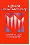
Light and Electron Microscopy - Elizabeth S. Slayter and Henry M. Slayter. The Slayter husband and wife team describes the principles of both electron and optical microscopy in terms of their applications in the biomedical and materials sciences. Included are discussions of image formation, contrast enhancement, and the ultimate limits on the size of observable details (resolving power and resolution), as well as a detailed account of Fourier optical theory. Photomicrography in electron and light microscopy is also reviewed, and an introduction to image processing techniques is clearly presented. Chapters on interference, polarized light, X-ray microscopy, scanning techniques, and innovative new concepts in microscope development offer excellent introductions to these important topics, as well. A detailed series of appendices on image location provides sign conventions, thin and thick lens equations, and ray-tracing techniques.
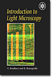
Introduction to Light Microscopy - Savile Bradbury and Brain Bracegirdle. Produced in association with the Royal Microscopical Society of London, this introductory volume starts with the basics of light and optics, and then proceeds into detailed discussions of microscope configuration. Simple and compound microscopes (including stereomicroscopes) are described in detail, as are their critical components, including eyepieces, objectives, condensers, and light sources. A considerable effort is made to delineate the important considerations in specimen illumination, primarily with modern Köhler techniques, but this volume also discusses older source-focused techniques, which are now becoming popular with light emitting diode microscopes. The last chapter on practical use of the microscope reviews basic concepts of imaging, low and high power objectives, oil immersion, darkfield, reflected light illumination, lamp adjustment, contrast enhancement, and microscope care and maintenance.
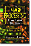
The Image Processing Handbook - John C. Russ. As digital imaging techniques become increasingly more popular (and necessary) in the field of optical microscopy, a thorough knowledge of the electronic processing methodology of images captured in the microscope becomes mandatory for students and researchers. Rapidly evolving technologies are reviewed in the latest edition of this best-selling volume, which emphasizes image measurements and includes new material on solid state cameras, compression algorithms, and printing innovations. The book also features information on image restoration, fuzzy logic in image processing, and applications in biology and optical microscopy. Extensive discussions of image restoration techniques, deblurring, and filtering algorithms are applied to both high-end graphics packages and popular desktop image-editing programs. A new chapter on stereology aids in quantitative analysis of biological specimens. This book should be considered an essential element for anyone studying digital imaging with the microscope.
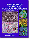
Handbook of Biological Confocal Microscopy - James B. Pawley (editor). With numerous contributions from leading researchers in the field, this second edition is a compendium of excellent review articles covering virtually all of the important aspects of laser scanning confocal microscopy. Included are reviews of light sources (both coherent and incoherent), objectives, optical systems, detectors, image processing, optical sectioning, deconvolution, reflection confocal, fluorophores, image contrast, specimen preservation, imaging living cells, multiphoton, lifetime imaging, and real time imaging. An extensive bibliography is also provided, although it is now somewhat dated. The appendices of the text contain useful diagrams of the scanning heads offered by major commercial manufacturers, and several articles contain information about the fundamental aspects of resolution and imaging in optical microscopy. A new volume is in the works, but this edition (published in 1995) is still available and contains one of the best collections of review articles available in a single location.
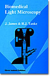
Biomedical Light Microscopy - J. James and H. J. Tanke. Targeted primarily at bioscience investigators, this introductory volume has been a favorite reference of microscopists for the past 25 years. The basic concepts of optical microscopy are discussed in detail, with strong emphasis being placed on the important topics of illumination, image formation, and resolution. An introductory chapter on fluorescence microscopy offers an excellent review of the numerous specimen considerations and the complex techniques involved, as well as a description of filter characteristics. Specialized techniques, such as phase contrast, interference contrast, modulation contrast, polarized light, and reflected light are briefly described. In addition, chapters on quantitative analysis and pattern recognition help introduce these important concepts to researchers in the field of cell biology. Although the chapter on photomicrography is somewhat dated, it should serve as a useful guide to those microscopists who are still using film camera systems.
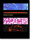
Plant Microtechnique and Microscopy - Steven E. Ruzin. Dr. Ruzin reviews the important field of microtechnique and provides an excellent working manual for students and researchers alike. Traditional and innovative new methodology is reviewed in step-by-step protocols, complete with a theoretical background to provide students with the necessary tools required to design and interpret their own experiments. Included are discussions of methacrylate embedding and sectioning, microwave tissue processing, fluorescence histochemistry, and in situ hybridization. The extensive appendices of the text provide common calculations, chemical toxicities, and buffer preparation tables. An excellent chapter on optical microscopy describes optical principles of techniques such as phase contrast, differential interference contrast (DIC), confocal, and deconvolution widefield microscopy. The appendix to the chapter includes a beneficial review of optical concepts as they apply to microscopy. Containing over 650 references, the bibliography provided with the text is a substantial resource for any student of histology or microscopy.
BACK TO MOLECULAR EXPRESSIONS MICROSCOPY PRIMER
