
Brightfield Microscopy Digital Image Gallery
Mammalian Cardiac Muscle Tissue
Muscle is the contractile tissue of animals, which functions in the production of both voluntary and involuntary movements. The three basic types of muscle are striated, smooth, and cardiac.
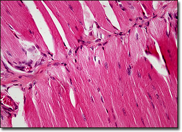
Comprised of elongated cells with multiple nuclei, cardiac muscle tissue appears striated under the microscope. Yet, unlike other striated muscles in the body, cardiac muscle controls an involuntary action, similar to smooth muscle tissue. The rhythmically contracting cardiac muscle tissue is essentially under the control of the heart’s pacemaker, the sinoatrial node. However, a number of chemical substances may affect the action of the tissue, many of which are utilized for medical purposes.
Referred to as inotropic agents, drugs that influence cardiac muscle may have a positive or negative effect, depending upon whether they increase or decrease the amount of force with which the heart contracts. Cardiac glycosides, which were utilized and somewhat understood as early as 1785, are generally believed to be the most important of all inotropic agents. Found in the leaves of various plants, cardiac glycosides increase the force of heart contractions and can, therefore, be utilized to treat heart failure. The substances are also associated, however, with a number of side effects, the most serious of which is ectopic heartbeats that can disrupt the rhythm of heart and may have fatal consequences.
BACK TO THE BRIGHTFIELD MICROSCOPY IMAGE GALLERY