
Brightfield Microscopy Digital Image Gallery
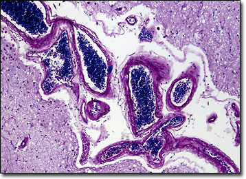
Human Cerebral Cortex
The human cerebrum is divided into two mirror image cerebral hemispheres by the longitudinal fissure. Each of these hemispheres exhibits an outer, wrinkly layer of gray matter known as the cerebral cortex.
In humans, the cerebral cortex is significantly larger than in other animals of similar size. Typically one to four millimeters thick with an approximate surface area of 2,000 square centimeters, the only way the sizable region of the brain can fit into the human skull is by folding extensively. The convolutions of the cerebral cortex are usually referred to in terms of sulci and gyri, which are respectively fissures and crests along the surface of the brain. The sulci and gyri exhibit patterns along the cerebral cortex and are the basis of the division of each hemisphere into six lobes. These lobes, which are usually identified as the frontal, parietal, temporal, occipital, central, and limbic lobes, are associated with various functions within the body.
The largest lobe of the cerebral cortex is the frontal lobe. A region of this lobe is sometimes referred to as Broca’s area, which is well known for its association with the ability to convert thoughts into words. The parietal lobe, however, is more commonly associated with motor function and the reception and integration of various kinds of sensory information from other parts of the body, while the temporal lobe is specifically related to hearing and the sense of smell, and the occipital lobe is heavily involved in proper vision. The central lobe, on the other hand, is not typically associated with any specific function, but the limbic lobe is generally believed to be involved in autonomic and visceral activities.
BACK TO THE BRIGHTFIELD MICROSCOPY IMAGE GALLERY