Brightfield Microscopy Digital Image Gallery
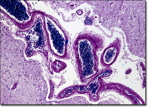
Human Cerebral Cortex
|
In humans, the cerebral cortex is significantly larger than in other animals of similar size. Typically one to four millimeters thick with an approximate surface area of 2,000 square centimeters, the only way the sizable region of the brain can fit into the human skull is by folding extensively. The convolutions of the cerebral cortex are usually referred to in terms of sulci and gyri, which are respectively fissures and crests along the surface of the brain. The sulci and gyri exhibit patterns along the cerebral cortex and are the basis of the division of each hemisphere into six lobes. These lobes, which are usually identified as the frontal, parietal, temporal, occipital, central, and limbic lobes, are associated with various functions within the body. |
© 1995-2025 by Michael W. Davidson and The Florida State University. All Rights Reserved. No images, graphics, software, scripts, or applets may be reproduced or used in any manner without permission from the copyright holders. Use of this website means you agree to all of the Legal Terms and Conditions set forth by the owners.
This website is maintained by our
|