
Chromatin and Chromosomes
Packed inside the nucleus of every human cell is nearly 6 feet of DNA, which is subdivided into 46 individual molecules, one for each chromosome and each about 1.5 inches long. Collecting all this material into a microscopic cell nucleus is an extraordinary feat of packaging. For DNA to function when necessary, it can't be haphazardly crammed into the nucleus or simply wound up like a ball of string. Consequently, during interphase, DNA is combined with proteins and organized into a precise, compact structure, a dense string-like fiber called chromatin, which condenses even further into chromosomes during cell division.
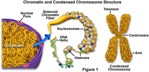
Each DNA strand wraps around groups of small protein molecules called histones, forming a series of bead-like structures, called nucleosomes, connected by the DNA strand (as illustrated in Figure 1). Under the microscope, uncondensed chromatin has a "beads on a string" appearance. The string of nucleosomes, already compacted by a factor of six, is then coiled into an even denser structure known as a solenoid that compacts the DNA by a factor of 40. The solenoid structure then coils to form a hollow tube. This complex compression and structuring of DNA serves several functions. The overall negative charge of the DNA is neutralized by the positive charge of the histone molecules, the DNA takes up much less space, and inactive DNA can be folded into inaccessible locations until it is needed.
There are two basic types of chromatin. Euchromatin is the genetically active type of chromatin involved in transcribing RNA to produce proteins used in cell function and growth. The predominant type of chromatin found in cells during interphase, euchromatin is more diffuse than the other kind of chromatin, which is termed heterochromatin. The additional compression of heterochromatin is thought to involve various proteins in addition to the histones, and the DNA it contains is thought to be genetically inactive. Heterochromatin tends to be most concentrated along chromosomes at certain regions of the structures, such as the centromeres and telomeres. Genes typically located in euchromatin can be experimentally silenced (not expressed) by relocating them to a heterochromatin position.
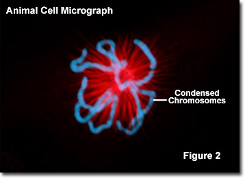
Throughout the life of a cell, chromatin fibers take on different forms inside the nucleus. During interphase, when the cell is carrying out its normal functions, the chromatin is dispersed throughout the nucleus in what appears to be a tangle of fibers. This exposes the euchromatin and makes it available for the transcription process. When the cell enters metaphase and prepares to divide, the chromatin changes dramatically. First, all the chromatin strands make copies of themselves through the process of DNA replication. Then they are compressed to an even greater degree as they undergo a 10,000-fold compaction into specialized structures for reproduction, the chromosomes (see Figure 2). As the cell divides to become two cells, the chromosomes separate, giving each cell a complete copy of the genetic information contained in the chromatin.
The number of chromosomes within the nuclei of an organism's cells is a species-specific trait. Human diploid cells (those that are not gametes) characteristically exhibit 46 chromosomes, but this number can be as low as 2, as is the case for some ants and roundworms, or more than a thousand, as exemplified by the Indian fern (Ophioglossum reticulatum), which has 1,260 chromosomes. Accordingly, the number of chromosomes a species has does not correlate to the complexity of the organism.