
The Endoplasmic Reticulum
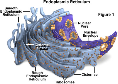
The endoplasmic reticulum (ER) is a network of flattened sacs and branching tubules that extends throughout the cytoplasm in plant and animal cells. These sacs and tubules are all interconnected by a single continuous membrane so that the organelle has only one large, highly convoluted and complexly arranged lumen (internal space). Usually referred to as the endoplasmic reticulum cisternal space, the lumen of the organelle often takes up more than 10 percent of the total volume of a cell. The endoplasmic reticulum membrane allows molecules to be selectively transferred between the lumen and the cytoplasm, and since it is connected to the double-layered nuclear envelope, it further provides a pipeline between the nucleus and the cytoplasm.
The endoplasmic reticulum manufactures, processes, and transports a wide variety of biochemical compounds for use inside and outside of the cell. Consequently, many of the proteins found in the cisternal space of the endoplasmic reticulum lumen are there only transiently as they pass on their way to other locations. Other proteins, however, are targeted to constantly remain in the lumen and are known as endoplasmic reticulum resident proteins. These special proteins, which are necessary for the endoplasmic reticulum to carry out its normal functions, contain a specialized retention signal consisting of a specific sequence of amino acids that enables them to be retained by the organelle. An example of an important endoplasmic reticulum resident protein is the chaperone protein known as BiP (formally: the chaperone immunoglobulin-binding protein), which identifies other proteins that have been improperly built or processed and keeps them from being sent to their final destinations.
There are two basic kinds of endoplasmic reticulum morphologies: rough and smooth. The surface of rough endoplasmic reticulum is covered with ribosomes, giving it a bumpy appearance when viewed through the microscope. This type of endoplasmic reticulum is involved mainly with the production and processing of proteins that will be exported, or secreted, from the cell. The ribosomes assemble amino acids into protein units, which are transported into the rough endoplasmic reticulum for further processing. These proteins may be either transmembrane proteins, which become embedded in the membrane of the endoplasmic reticulum, or water-soluble proteins, which are able to pass completely through the membrane into the lumen. Those that reach the inside of the endoplasmic reticulum are folded into the correct three-dimensional conformation, as a flattened cardboard box might be opened up and folded into its proper shape in order to become a useful container. Chemicals, such as carbohydrates or sugars, are added, then the endoplasmic reticulum either transports the completed proteins to areas of the cell where they are needed, or they are sent to the Golgi apparatus for further processing and modification.
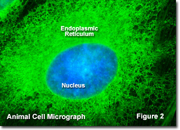
Most proteins exported from the endoplasmic reticulum exit the organelle in vesicles budded from the smooth portion, which has a more even appearance than rough endoplasmic reticulum when viewed through the electron microscope because of the lack of ribosomes. The smooth endoplasmic reticulum in most cells is much less extensive than the rough endoplasmic reticulum and is sometimes alternatively termed transitional. Smooth endoplasmic reticulum is chiefly involved, however, with the production of lipids (fats), building blocks for carbohydrate metabolism, and the detoxification of drugs and poisons. Therefore, in some specialized cells, such as those that are occupied chiefly in lipid and carbohydrate metabolism (brain and muscle) or detoxification (liver), the smooth endoplasmic reticulum is much more extensive and is crucial to cellular function. Smooth endoplasmic reticulum also plays a role in various cellular activities through its storage of calcium and involvement in calcium metabolism. In muscle cells, smooth endoplasmic reticulum releases calcium to trigger muscle contractions. Presented in Figure 2 is a fluorescence digital image taken through the microscope of the endoplasmic reticulum network in a bovine (cow) pulmonary artery endothelial cell grown in culture.
It is the rough endoplasmic reticulum that is directly continuous with the nuclear envelope (as illustrated in Figure 1), which is also studded with ribosomes, and the two organelles are thought to have evolved simultaneously in ancient cells. Due to their physical membranous connection, the lumen of the endoplasmic reticulum and the space between the layers of the nuclear envelope comprise a single compartment. Accordingly, the nucleus has direct access to proteins (many of which are produced by the ribosomes upon its surface) and other materials present in the endoplasmic reticulum lumen, so that transport vesicles are not needed to obtain them. The close association between the endoplasmic reticulum and the nucleus also enables the organelles to share information in a very efficient manner. For instance, if the endoplasmic reticulum begins to undergo functional problems and unfolded proteins accumulate within the organelle, which can be extremely hazardous to the cell, the organelle quickly sends a signal to the nucleus (as well as to the cytoplasm). The nucleus responds by slowing ribosomal translation through a several-step process, thereby giving the endoplasmic reticulum extra time to catch up on its protein folding, thus maintaining cellular health.